IPLab:Lab 7:Metastatic Adenocarcinoma
Jump to navigation
Jump to search
Clinical Summary
This 58-year-old male was admitted five weeks earlier with a weight loss of 80 pounds over a six-month period, abdominal cramps, and rebound tenderness in the right lower quadrant. Abdominal and chest imaging showed multiple nodular masses in the lungs and liver. Fine needle biopsy of the liver revealed adenocarcinoma with the primary source thought to be colon. He was discharged on chemotherapy, but returned two days later with small bowel obstruction and sepsis, and he died a few days later.
Autopsy revealed an obstructive firm mass in the cecum with similar masses in the lungs, lymph nodes, liver and peritoneum. A large retrocecal abscess was found. Blood cultures grew Klebsiella pneumoniae and E. coli.
Images
-
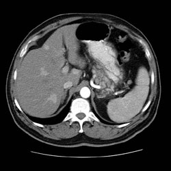 Abdominal CT showing multiple nodular masses in the liver.
 -
 This gross photograph of the liver from this case demonstrates multiple, variably-sized pale/white-tan nodules scattered throughout the liver.
 -
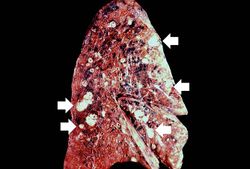 This gross photograph of the lung from this case also demonstrates multiple, variably sized pale/white-tan nodules scattered throughout the lung.
 -
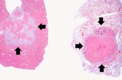 These are low-power photomicrographs of a section of liver (left) and lung (right) containing tumor nodules (arrows).
 -
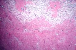 This is a photomicrograph taken at the interface between the tumor (top) and the normal liver parenchyma (bottom).
 -
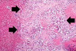 This is a higher-power photomicrograph showing how the tumor cells (arrows) have infiltrated into the liver parenchyma.
 -
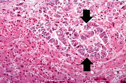 This is a high-power photomicrograph of tumor cells that are forming glands (arrows).
 -
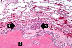 This is a photomicrograph of a tumor nodule in the lung. The tumor cells are infiltrating into the lung parenchyma (1). There is a large area of necrosis in the center of the tumor (2).
 -
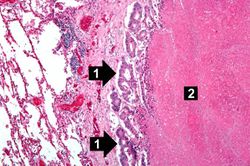 This is a high-power photomicrograph of the edge of the tumor nodule in the lung. The tumor cells are infiltrating into the lung parenchyma (1). Even at this power you can see the glandular formation of this adenocarcinoma. There is a large area of necrosis in the center of the tumor (2).
 -
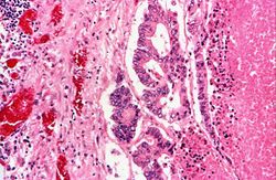 This is a high-power photomicrograph of the edge of the tumor nodule in the lung. The tumor cells area growing in a glandular pattern. The area of necrosis is evident at the right side of the image.


Virtual Microscopy
Lung
Liver
Study Questions
Additional Resources
Reference
- eMedicine Medical Library: Imaging in Adenocarcinoma of the Colon
- eMedicine Medical Library: Colon Adenocarcinoma
- Merck Manual: Colorectal Cancer
- Merck Manual: Metastatic Liver Cancer
Journal Articles
- Castro CY, Moran CA, Flieder DG, Suster S. Primary signet ring cell adenocarcinomas of the lung: a clinicopathological study of 15 cases. Histopathology 2001 Oct;39(4):397-401.
Images
- PEIR Digital Library: Adenocarcinoma Images
- PEIR Digital Library: Metastatic Adenocarcinoma Images
- WebPath: Hepatic Neoplasms
- WebPath: Lung Neoplasms
Related IPLab Cases
- Lab 7: Colon: Adenocarcinoma
- Lab 7: Lip: Squamous Cell Carcinoma
- Lab 7: Esophagus: Squamous Cell Carcinoma
- Lab 7: Breast: Infiltrating Ductal Carcinoma
- Lab 7: Lung: Bronchogenic Carcinoma