IPLab:Lab 7:Carcinoid
Jump to navigation
Jump to search
Clinical Summary
This 58-year-old male experienced increasing diarrhea over the 4 months prior to admission. During this period he experienced a weight loss of 40 pounds. Imaging demonstrated a lesion a the ileocecal valve and a laparotomy was performed.
The operative specimen consisted of 12 cm of distal ileum, appendix, cecum and colon. On opening the bowel there was a 4.5 x 3 x 3-cm elliptical submucosal mass at the ileocecal valve that had produced partial obstruction. Several small (2 mm) submucosal masses were found in the cecum nearby. On cut section each lesion was found to be firm, gray-tan and homogeneous involving the muscular wall of the bowel and adjacent mesentery.
Images
-
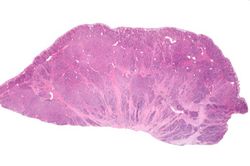 This is a low-power photomicrograph of the surgical specimen showing basophilic and eosinophilic areas delimiting areas of tumor infiltration.
 -
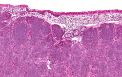 This is a higher-power photomicrograph of the surgical specimen showing the mucosa is normal and the tumor cells are in the submucosa.
 -
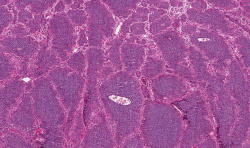 This is a higher-power photomicrograph of the surgical specimen showing nests of tumor cells.
 -
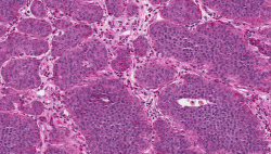 This is a high-power photomicrograph of the surgical specimen showing the tumor's growth pattern--cells form discrete islands, trabeculae, and glands.
 -
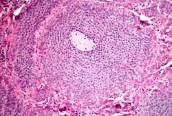 This is a high-power photomicrograph of the surgical specimen showing the cellular morphology. The tumor cells are monotonously similar with scant, pink, granular cytoplasm and a round-to-oval stippled nucleus. As in most carcinoid tumors, there is minimal variation in cell and nuclear size, and mitoses are infrequent or absent.
 -
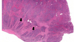 This low-power photomicrograph shows how the tumor cells have invaded into the muscularis of the bowel (arrows).


Virtual Microscopy
Study Questions
Additional Resources
Reference
- eMedicine Medical Library: Malignant Carcinoid Syndrome
- Merck Manual: Overview of Carcinoid Tumors
- Merck Manual: Carcinoid Syndrome
Journal Articles
- Shebani KO, Souba WW, Finkelstein DM, Stark PC, Elgadi KM, Tanabe KK, Ott MJ. Prognosis and survival in patients with gastrointestinal tract carcinoid tumors. Ann Surg 1999 Jun;229(6):815-21; discussion 822-3.