IPLab:Lab 7:Malignant Melanoma: Difference between revisions
Jump to navigation
Jump to search
Seung Park (talk | contribs) |
Seung Park (talk | contribs) No edit summary |
||
| Line 13: | Line 13: | ||
File:IPLab7Melanoma8.jpg|This is a high-power photomicrograph of the main tumor mass showing the cellular details. The individual melanoma cells contain large nuclei with irregular contours having chromatin clumped at the periphery of the nuclear membrane and prominent red (eosinophilic) nucleoli. | File:IPLab7Melanoma8.jpg|This is a high-power photomicrograph of the main tumor mass showing the cellular details. The individual melanoma cells contain large nuclei with irregular contours having chromatin clumped at the periphery of the nuclear membrane and prominent red (eosinophilic) nucleoli. | ||
</gallery> | </gallery> | ||
== Virtual Microscopy == | |||
<peir-vm>IPLab7Melanoma</peir-vm> | |||
== Study Question == | == Study Question == | ||
Revision as of 11:25, 3 January 2014
Clinical Summary
This 68-year-old white male had a local excision of a pigmented lesion (melanoma) on the skin of his back. Three years later he became aware of a "lump" in his left axilla. Examination confirmed the presence of a 2.3-cm nodular lesion. Subsequently, the patient underwent a surgical procedure for removal of axillary lymph nodes.
Images
-
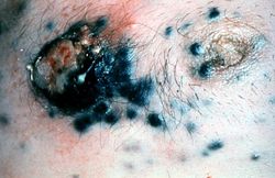 This is a gross photograph of skin with melanoma. Note the black pigment, multiple satellite nodules, and focal ulceration. Some of the satellite nodules affect the nipple.
 -
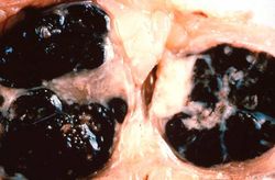 This is a gross photograph of lymph nodes almost entirely replaced by black pigment (melanin).
 -
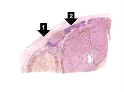 This is a low-power photomicrograph of lymph node that is almost completely replaced/filled with tumor. This lymph node has a capsule (1) and some remaining lymphocytes (2) but the remainder of the node is replaced by tumor cells.
 -
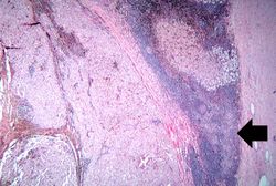 This higher-power photomicrograph shows the remaining portion of lymph node (arrow). The rest of the lymph node is invaded by a neoplasm composed of cells with lighter eosinophilic cytoplasm and pigment.
 -
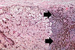 This is a higher-magnification showing the abundant extracellular melanin (arrows) surrounding the tumor cells. This section of neoplasm shows the numerous cells with abundant cytoplasm and brown pigment within the cytoplasm of some of these cells.
 -
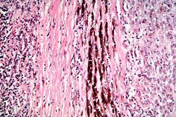 This is a higher magnification showing the abundant extracellular melanin surrounding the tumor cells (brown pigment).
 -
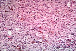 This is a high-power photomicrograph of the main tumor mass with the cells growing as poorly formed nests and sheets of cells. There is little if any pigment in this section.
 -
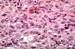 This is a high-power photomicrograph of the main tumor mass showing the cellular details. The individual melanoma cells contain large nuclei with irregular contours having chromatin clumped at the periphery of the nuclear membrane and prominent red (eosinophilic) nucleoli.


Virtual Microscopy
Study Question
Additional Resources
Reference
- eMedicine Medical Library: Cutaneous Melanoma
- eMedicine Medical Library: Malignant Melanoma
- eMedicine Medical Library: Skin Cancer -- Melanoma
- Merck Manual: Melanoma
Journal Articles
- Banerjee SS, Harris M. Morphological and immunophenotypic variations in malignant melanoma. Histopathology 2000 May;36(5):387-402.