IPLab:Lab 7:Adenoma: Difference between revisions
Jump to navigation
Jump to search
Seung Park (talk | contribs) No edit summary |
Seung Park (talk | contribs) |
||
| Line 27: | Line 27: | ||
== Additional Resources == | == Additional Resources == | ||
=== Reference === | === Reference === | ||
* [http://emedicine.medscape.com/article/850823-overview eMedicine Medical Library: Evaluation of Solitary Thyroid Nodule] | |||
* [http://emedicine.medscape.com/article/127491-overview eMedicine Medical Library: Thyroid Nodule] | |||
=== Journal Articles === | === Journal Articles === | ||
Revision as of 14:06, 23 August 2013
Clinical Summary
This 80-year-old white female's death came as the result of cardiopulmonary disease -- hypertension, coronary artery disease, pulmonary emphysema and cardiac hypertrophy.
Autopsy Findings
During a routine postmortem examination, this patient's thyroid gland was found to be nodular. The right lobe contained several colloid nodules. Located in the left lobe was a 2-cm well-circumscribed mass.
Images
-
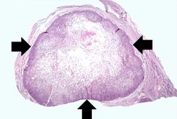 This is a low-power photomicrograph of a nodule found in the thyroid of this case. Note that the mass is well-circumscribed and there is a sharp line of demarcation between the mass and the adjacent thyroid tissue (arrows).
 -
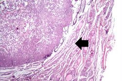 This is a higher-power view of the border between the tumor mass and the adjacent thyroid tissue. Note that the mass has compressed the adjacent normal thyroid tissue (arrow). Also note the different morphology between the adenoma (very cellular, dense follicles, little or no colloid) and the adjacent normal thyroid (larger follicles, colloid).
 -
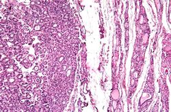 This is another higher-power photomicrograph of the adenoma (left) and the adjacent thyroid tissue (right). Note the compression of the adjacent normal thyroid and the difference in morphology between the adenoma and the thyroid.
 -
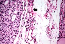 This photomicrograph demonstrates the densely packed follicular pattern in the adenoma (left) and the larger colloid-filled follicles of the normal thyroid (right). An area of compressed thyroid is present adjacent to the adenoma (arrows).
 -
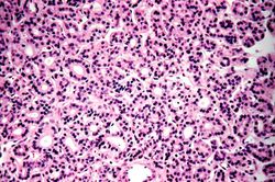 This is a photomicrograph of an adenoma. Note that the follicular architecture is well developed and more or less uniform throughout this section.
 -
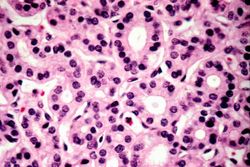 This high-power photomicrograph demonstrates the relatively normal cellular morphology of this follicular adenoma.


Study Questions
Additional Resources
Reference
- eMedicine Medical Library: Evaluation of Solitary Thyroid Nodule
- eMedicine Medical Library: Thyroid Nodule