IPLab:Lab 7:Esophagus SCC: Difference between revisions
Seung Park (talk | contribs) No edit summary |
Seung Park (talk | contribs) No edit summary |
||
| Line 29: | Line 29: | ||
* genetic (racial) predisposition.</spoiler> | * genetic (racial) predisposition.</spoiler> | ||
* <spoiler text="What factors facilitate growth and spread of this tumor?">The rich lymphatic network in the submucosa of the esophagus promotes extensive circumferential and longitudinal spread. Intramural tumor cell clusters are often seen several centimeters away from the main tumor mass. Local extension into adjacent mediastinal structures occurs early and often in this disease and seriously limits the chance of curative resection.</spoiler> | * <spoiler text="What factors facilitate growth and spread of this tumor?">The rich lymphatic network in the submucosa of the esophagus promotes extensive circumferential and longitudinal spread. Intramural tumor cell clusters are often seen several centimeters away from the main tumor mass. Local extension into adjacent mediastinal structures occurs early and often in this disease and seriously limits the chance of curative resection.</spoiler> | ||
== Additional Resources == | |||
=== Reference === | |||
=== Journal Articles === | |||
=== Images === | |||
== Related IPLab Cases == | |||
{{IPLab 7}} | {{IPLab 7}} | ||
[[Category: IPLab:Lab 7]] | [[Category: IPLab:Lab 7]] | ||
Revision as of 23:20, 22 August 2013
Clinical Summary
Approximately six months prior to admission, this 78-year-old male began having difficulty in swallowing solid food. This difficulty was described as a sticking of the food in his throat and was accompanied by cramping pain which could only be relieved by "coughing up" the ingested food. This dysphagia was accompanied by a twenty-pound weight loss. Following an upper GI series and endoscopic biopsy, the patient was given radiation treatment with considerable improvement. He did well for four months, after which the dysphagia and weight loss increased markedly. He refused operative intervention or further treatment and he died at home two months later.
Autopsy Findings
An autopsy revealed a circumferential fungating mass in the distal third of the esophagus. This mass partially occluded the lumen of the esophagus.
Images
-
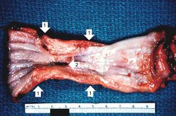 This is a gross photograph of the luminal surface of the esophagus with the area of constriction (1). The area protrudes into the lumen. There is also a central area of ulceration (2).
 -
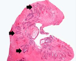 This low-power photomicrograph of a cross-section through the esophagus at the area of constriction shows extensive infiltration of the esophageal wall with squamous cell carcinoma (arrows).
 -
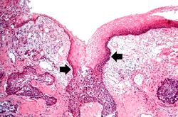 This is a high-power photomicrograph demonstrating the normal epithelium undergoing transition to carcinoma (arrows).
 -
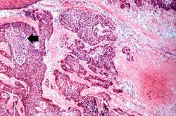 This is a higher-power photomicrograph showing invasive squamous cell carcinoma. Tongues and islands of tumor cells exhibit areas of central necrosis (arrow).
 -
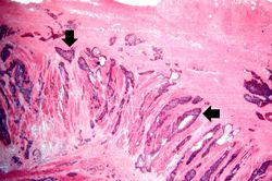 This is a photomicrograph of bands of tumor cells invading into the adjacent tissues (arrows).
 -
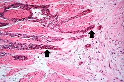 This is a higher-power photomicrograph of bands of tumor cells (arrows) extending between the muscle bundles.
 -
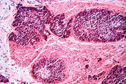 This is a high-power photomicrograph of the tumor cells that have invaded the adjacent muscle tissue.


