IPLab:Lab 7:Osteosarcoma: Difference between revisions
| Line 3: | Line 3: | ||
The distal diaphysis of the femur and adjacent soft tissues were involved in a 15 x 10 x 10-cm mass. The cut surface of the mass was fleshy white, with focal areas of hemorrhage. | The distal diaphysis of the femur and adjacent soft tissues were involved in a 15 x 10 x 10-cm mass. The cut surface of the mass was fleshy white, with focal areas of hemorrhage. | ||
== Images == | == Images == | ||
Revision as of 20:49, 9 July 2020
Clinical Summary
This 14-year-old male first experienced mild pain in the left knee after playing baseball, approximately two months ago. The pain persisted in an intermittent fashion until approximately two weeks prior to admission, when the pain increased significantly and was accompanied by marked swelling and loss of motion. He also had decreased appetite, lethargy, and a 10-pound weight loss. On physical examination, the left knee was enlarged diffusely, firm, and non-tender. Radiographic and biopsy results confirmed a diagnosis of osteosarcoma. The patient underwent surgical removal of the distal femur and knee with placement of a prosthetic knee joint and bone grafts.
The distal diaphysis of the femur and adjacent soft tissues were involved in a 15 x 10 x 10-cm mass. The cut surface of the mass was fleshy white, with focal areas of hemorrhage.
Images
-
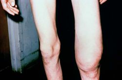 This is a photograph of the patient prior to surgery. Note the marked swelling of the knee.
 -
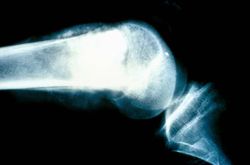 This is a radiograph showing the tumor in the distal femur.
 -
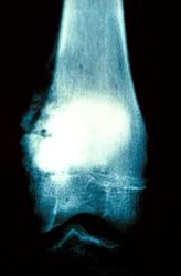 This is another view of the tumor in the distal femur.
 -
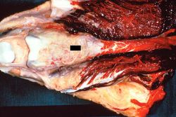 This is a gross photograph of the surgical specimen with tissue dissected away to demonstrate the tumor mass.
 -
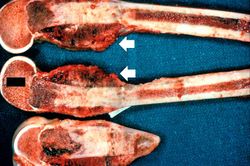 These are cut sections of the distal femur containing the tumor. The periosteal involvement is evident from this picture (arrows).
 -
 This is a low-power photomicrograph of decalcified histologic section from this tumor. Note the blue color (cell nuclei stain blue) of much of this section indicating the increased cellularity of the tumor.
 -
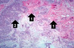 This is a higher-power photomicrograph of decalcified histologic section from this tumor. There are areas of osteoid (1) and cellular areas (2).
 -
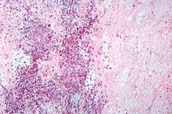 This is a high-power photomicrograph of decalcified histologic section showing the cellularity of the tumor.
 -
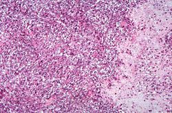 This high-power photomicrograph demonstrates the cellular growth pattern. Note that the cells are fusiform and they grow in sheets.
 -
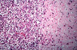 This high-power photomicrograph demonstrates the growth pattern and the cell morphology.
 -
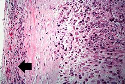 This is a high-power photomicrograph of the tumor cell morphology and the periosteum (arrow).
 -
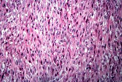 This high-power photomicrograph of the tumor demonstrates the fusiform morphology of the cells. Note the marked variability in size and staining intensity of the nuclei.
 -
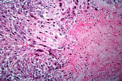 This is a high-power photomicrograph of the tumor demonstrating the anaplastic cell morphology.
 -
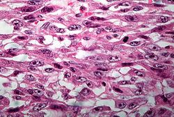 This is a high-power photomicrograph of the tumor demonstrating the anaplastic cell morphology.
 -
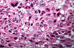 This is a high-power photomicrograph of the tumor demonstrating the anaplastic cell morphology and multiple mitotic figures (arrows).


Virtual Microscopy
<peir-vm>IPLab7Osteosarcoma</peir-vm>
Study Question
- <spoiler text="What is the incidence and age distribution of osteosarcomas?">Osteosarcoma is the most common primary malignant tumor of bone exclusive of myeloma and lymphoma and accounts for approximately 20% of primary bone cancers. Males are more commonly affected than females (1.6:1). Osteosarcoma occurs in all age groups but has a bimodal age distribution. Approximately 75% occur in patients younger than 20 years of age. The smaller second peak (secondary osteosarcoma) occurs in elderly patients who suffer from conditions known to be associated with the development of osteosarcoma (Paget’s disease, bone infarcts, and prior irradiation).</spoiler>
- <spoiler text="What are the most common locations for osteosarcomas?">Osteosarcomas usually arise in the metaphyseal region of the long bones of the extremities, and almost 60% occur about the knee. The location, in descending order of frequency, is distal femur, proximal tibia, proximal humerus, and proximal femur.</spoiler>
- <spoiler text="What are some of the risk factors associated with the pathogenesis of osteosarcoma?">Gene mutations are fundamental to the development of osteosarcoma. Patients with hereditary retinoblastomas have a several hundred-fold greater risk of developing osteosarcoma. Mutations in the p53 suppressor gene (Li-Fraumeni syndrome) has been implicated in the development of most nonhereditary osteosarcomas as well as many other types of cancer. Interestingly, many osteosarcomas develop at sites of greatest bone growth where bone cell mitotic activity is at its peak--e.g., the base of the femoral growth plate where primary spongiosa is being formed and in Pagetic bone with its frenzy of bone formation and resorption.</spoiler>
- <spoiler text="What is the prognosis for patients with osteosarcoma?">Osteosarcomas are aggressive neoplasms that spread through the bloodstream, and at the time of diagnosis, approximately 20% of patients have demonstrable pulmonary metastases. In those who die of the neoplasm, 90% have metastases to the lungs, bones, brain, and elsewhere. With current treatment regimens the long-term survival rate has been increased to approximately 60% from the historic controls of 25%.</spoiler>
Additional Resources
Reference
Journal Articles
- Wittig JC, Bickels J, Priebat D, Jelinek J, Kellar-Graney K, Shmookler B, Malawer MM. Osteosarcoma: a multidisciplinary approach to diagnosis and treatment. Am Fam Physician 2002 Mar 15;65(6):1123-32.
- Miller SL, Hoffer FA. Malignant and benign bone tumors. Radiol Clin North Am 2001 Jul;39(4):673-99.