Difference between revisions of "IPLab:Lab 7:Esophagus SCC"
Seung Park (talk | contribs) (Created page with "== Images == <gallery heights="250px" widths="250px"> File:IPLab7EsophSCC1.jpg|This is a gross photograph of the luminal surface of the esophagus with the area of constriction...") |
Seung Park (talk | contribs) |
||
| Line 1: | Line 1: | ||
| + | == Clinical Summary == | ||
| + | Approximately six months prior to admission, this 78-year-old male began having difficulty in swallowing solid food. This difficulty was described as a sticking of the food in his throat and was accompanied by cramping pain which could only be relieved by "coughing up" the ingested food. This dysphagia was accompanied by a twenty-pound weight loss. Following an upper GI series and endoscopic biopsy, the patient was given radiation treatment with considerable improvement. He did well for four months, after which the dysphagia and weight loss increased markedly. He refused operative intervention or further treatment and he died at home two months later. | ||
| + | |||
| + | == Autopsy Findings == | ||
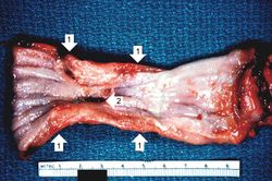
| + | An autopsy revealed a circumferential fungating mass in the distal third of the esophagus. This mass partially occluded the lumen of the esophagus. | ||
| + | |||
== Images == | == Images == | ||
<gallery heights="250px" widths="250px"> | <gallery heights="250px" widths="250px"> | ||
Revision as of 14:32, 21 August 2013
Clinical Summary[edit]
Approximately six months prior to admission, this 78-year-old male began having difficulty in swallowing solid food. This difficulty was described as a sticking of the food in his throat and was accompanied by cramping pain which could only be relieved by "coughing up" the ingested food. This dysphagia was accompanied by a twenty-pound weight loss. Following an upper GI series and endoscopic biopsy, the patient was given radiation treatment with considerable improvement. He did well for four months, after which the dysphagia and weight loss increased markedly. He refused operative intervention or further treatment and he died at home two months later.
Autopsy Findings[edit]
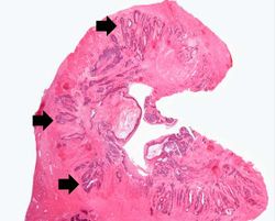
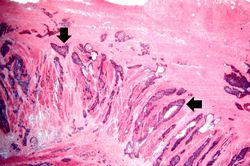
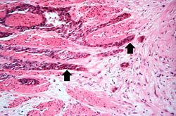
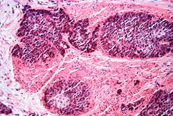
An autopsy revealed a circumferential fungating mass in the distal third of the esophagus. This mass partially occluded the lumen of the esophagus.
Images[edit]
An upper GI series is a series of barium-aided radiographs involving the esophagus, stomach, and duodenum.
