IPLab:Lab 13:Biliary Atresia
Clinical Summary
This eight-month-old female developed jaundice at one week of age. As the jaundice deepened, the child's bilirubin level was found to be over 30 mg/dL and the patient experienced a few convulsive episodes. A liver biopsy produced a diagnosis of extrahepatic bile duct obstruction. A subsequent exploratory laparotomy showed an absence of extrahepatic bile duct structures--including the gallbladder--and an intraoperative cholangiogram failed to show any large biliary channel. A jejunohepatostomy in the portal area was performed in the hope of establishing bile drainage and the patient was placed on the waiting list for a liver transplant. However, the patient progressively deteriorated, developed infections, and after a long turbulent course, died of pneumonia.
An autopsy was performed and the diagnosis of extrahepatic bile duct obstruction was confirmed.
Images
-
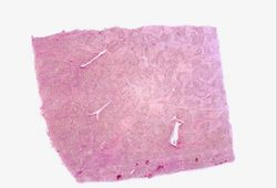 This is a low power photomicrograph of a section of liver from this stillborn infant. Even at this low magnification, areas of fibrosis can be appreciated.
 -
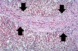 This medium-power photomicrograph of liver shows an area of portal fibrosis and bile duct proliferation (arrows). Adjacent to this fibrotic portal region, hepatocytes are seen separated by dilated sinusoids. Throughout this section are found accumulations of yellow-brown bile pigment.
 -
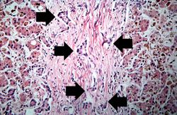 This high-power photomicrograph of fibrotic portal region demonstrates proliferation of the bile ducts (arrows).
 -
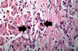 This is a high-power photomicrograph of fibrotic portal region with several bile ducts that contain inspissated bile (arrows). Adjacent hepatocytes also contain bile pigments.
 -
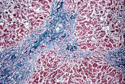 This is a medium-power photomicrograph of liver section stained with a trichrome stain to demonstrate the portal fibrosis. The fibrous connective tissue (collagen) stains blue.


Virtual Microscopy
H&E
Masson Trichrome
Study Questions
Additional Resources
Reference
Journal Articles
- Davenport M, Caponcelli E, Livesey E, Hadzic N, Howard E. Surgical outcome in biliary atresia: etiology affects the influence of age at surgery. Ann Surg 2008 Apr;247(4):694-8.
Images
Related IPLab Cases
| |||||