IPLab:Lab 5:Hemochromatosis
Jump to navigation
Jump to search
Clinical Summary
This 61-year-old female was first admitted to the hospital because of ascites and pedal edema. A liver biopsy revealed a marked intracellular accumulation of iron. The serum iron concentration was increased at 220 mcg/dL. On the basis of these studies, the diagnosis of hemochromatosis was made. Subsequent to the first admission, the patient was admitted on several occasions for ascites. The patient's last admission was necessitated by the development of symptoms and signs of hepatic failure characterized by jaundice and coma.
At autopsy the liver weighed 800 grams. The cut surface was described as golden-brown in color, having a fine, diffuse nodularity, and being extremely firm in consistency.
Images
-
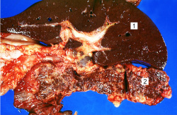 This is a gross photograph of liver (1) and pancreas (2) from this case of hemochromatosis. Note that both of these organs have a dark brown coloration.
 -
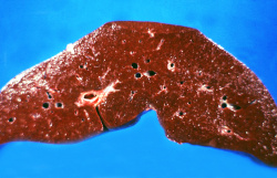 This is a gross photograph of a cut section of liver from this case of hemochromatosis. Note that the liver is dark brown. Although hard to appreciate in a photograph, the tissue is also firm (cirrhotic).
 -
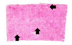 This is a low-power micrograph of liver from this patient. Note the nodularity of the tissue (arrows).
 -
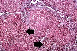 This higher-power view of liver from this case demonstrates the nodules and the brown/black pigment within liver parenchymal cells (arrows).
 -
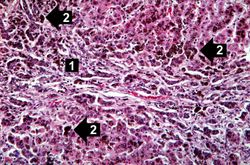 This higher-power photomicrograph demonstrates the increased fibrosis in the periportal area (1) and the pigment accumulation (2).
 -
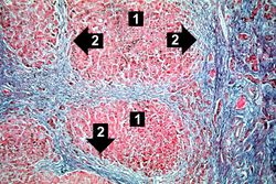 This trichrome stain of liver section demonstrates the increased fibrous connective tissue in this liver. Note that the liver nodules (1) are surrounded by fibrous connective tissue (2).
 -
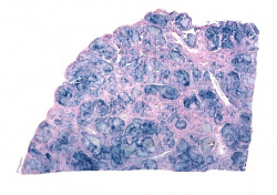 This is a low-power view of liver section stained with Prussian blue. Prussian blue reacts with iron in the tissue to give a blue color.
 -
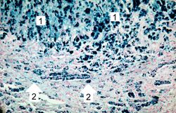 This higher-power view of liver stained with Prussian blue demonstrates the marked accumulation of iron within the parenchymal cells (1) and in the Kupffer cells in the periportal area (2).
 -
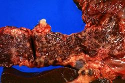 This is a gross picture of pancreas from this case. Note the brown discoloration of the tissue.
 -
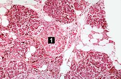 This is a histologic section of pancreas from this case. It is difficult to appreciate at this magnification, but there is brown pigment in the pancreatic acinar cells. Note the islets of Langerhans (1).
 -
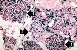 This is a histologic section of pancreas from this case stained for iron (Prussian blue). Note the accumulation of iron in the parenchymal cells (1). There is also iron in the pancreatic islets (2).


Virtual Microscopy
H&E
Liver: Hemochromatosis
Normal Liver
Prussian Blue
Study Questions
- Compare and contrast the pathogenesis and clinical features of hemochromatosis and Wilson’s disease.
Additional Resources
Reference
- eMedicine Medical Library: Hemochromatosis
- Merck Manual: Iron Overload: Hemosiderosis and Hemochromatosis
Journal Articles
- Powell LW, Seckington RC, Deugnier Y. Haemochromatosis. Lancet 2016.
- Ayonrinde OT, Milward EA, Chua AC, Trinder D, Olynyk JK. Clinical perspectives on hereditary hemochromatosis. Crit Rev Clin Lab Sci 2008;45(5):451-84.
- Bassett ML. Haemochromatosis: iron still matters. Intern Med J 2001 May-Jun;31(4):237-42.
Images
- PEIR Digital Library: Hemochromatosis Images
- WebPath: Hepatic Pigmentary Disorders
- WebPath: Cellular Accumulations
Related IPLab Cases
| |||||