IPLab:Lab 7:Fibroadenoma
Jump to navigation
Jump to search
Clinical Summary
Four months prior to admission, this 25-year-old female became aware of a lump beneath the areola of her right breast. Physical examination confirmed the presence of an approximately 3 cm movable, rubbery mass. An aspiration was attempted but did not yield any fluid or cells. At the time of surgical exploration, a well-circumscribed mass was identified and removed.
Images
-
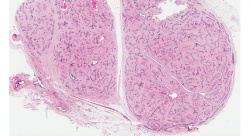 This low-power photomicrograph of the surgical specimen demonstrates two ovoid, well-circumscribed nodules surrounded by fibroadipose tissue.
 -
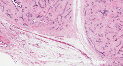 In this higher magnification you can see the solid parenchyma with numerous variable sized glandular spaces.
 -
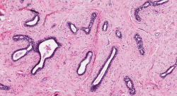 This is a higher magnification of the fibroadenoma showing the dense stroma of the tumor surrounding the irregularly shaped ducts.
 -
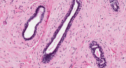 This is a high magnification of the fibroadenoma showing the dense stroma of the tumor surrounding the irregularly shaped ducts. The ducts are lined by two cell layers, one of cuboidal, two columnar cells (inner layer) and an outer layer of flattened cells with hyperchromatic nuclei (myoepithelial cells).


Virtual Microscopy
Study Questions
Additional Resources
Reference
Journal Articles
- Yilmaz E, Sal S, Lebe B. Differentiation of phyllodes tumors versus fibroadenomas. Acta Radiol 2002 Jan;43(1):34-9.
- Hogge JP, De Paredes ES, Magnant CM, Lage J. Imaging and management of breast masses during pregnancy and lactation. Breast J 1999 Jul;5(4):272-283.