IPLab:Lab 5:Trisomy 21
Clinical Summary
This 44-year-old woman was 12 weeks pregnant with her third child when routine blood work showed a maternal serum alpha-fetoprotein level that was lower than expected for her stage of pregnancy. At 15-weeks of gestation she underwent ultrasound-guided amniocentesis and samples of the fluid containing cells were smeared onto glass slides for fluorescent in situ hybridization (FISH). The remainder of the sample was spun down and the fluid was sent for analysis and the cells were cultured for karyotyping.
FISH analysis demonstrated three copies of chromosome 21. The patient was informed of these results and was told that definitive results from the karyotyping studies would take several days. The patient was referred to a medical geneticist for counseling.
Images
-
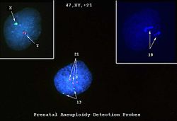 This is a photomicrograph of cells obtained by amniocentesis that were stained using FISH. The cell in panel 1 was stained with markers specific for the X and Y-chromosomes. The cell in panel 2 was stained with a marker specific for chromosome 18. The cell in the center was stained with markers for chromosomes 13 and 21. Note that there are three copies of chromosome 21.
 -
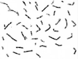 These cells, obtained by amniocentesis, were cultured and then arrested in metaphase. Nuclei from these cells were isolated and stained to demonstrate the banding pattern of each chromosome. This photograph shows a "chromosome spread." Each chromosome is identified and lined up to give a karyotype (next page).
 -
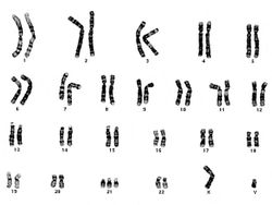 Chromosomes from the chromosome spread are lined up to demonstrate the karyotype. In this case there are three copies of chromosome 21, just as noted in the FISH.
 -
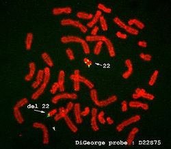 FISH is also useful in the diagnosis of other genetic disorders. This is an example of FISH staining on another patient using a probe specific for DiGeorge's disease. The arrow shows that there is a deletion on chromosome 22, which is diagnostic for DiGeorge's disease.
 -
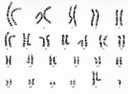 This is a karyotype of a patient with Klinefelter syndrome (47, XXY).
 -
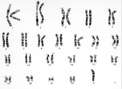 This is a karyotype of a patient with Turner syndrome (45, X).


Study Questions
Additional Resources
Reference
- eMedicine Medical Library: Down Syndrome
- eMedicine Medical Library: Klinefelter Syndrome
- eMedicine Medical Library: Turner Syndrome
- Merck Manual: Chromosomal Abnormalities
Journal Articles
- Garcia-Heras J, Rao PN. A brief review of cryptic duplications of 21q as an emerging cause of Down syndrome: practical considerations for accurate detection. Clin Genet 1999 Mar;55(3):207-11.
Images
- PEIR Digital Library: Down Syndrome Images
- PEIR Digital Library: Genetics Images
- WebPath: Pediatric-Perinatal Pathology
| |||||