Difference between revisions of "IPLab:Lab 6:Hashimoto's Thyroiditis"
(→Images) |
|||
| (13 intermediate revisions by 2 users not shown) | |||
| Line 1: | Line 1: | ||
| + | == Clinical Summary == | ||
| + | This was a 49-year-old woman who complained of tiredness and difficulty concentrating. She had gained weight over the last year and despite warm weather, she felt chilled without a sweater. Family history was significant for hypothyroidism in her mother and older sister. | ||
| + | |||
| + | On physical examination she had an enlarged thyroid gland with a firm, bosselated texture. Serum TSH was markedly elevated and antithyroid peroxidase antibodies were positive. These results supported the clinical impression of hypothyroidism; also, the texture of her thyroid gland and a positive family history suggested an autoimmune etiological factor. She was referred to an endocrinologist; however, before beginning treatment she died suddenly from a ruptured berry aneurysm. | ||
| + | |||
== Images == | == Images == | ||
<gallery heights="250px" widths="250px"> | <gallery heights="250px" widths="250px"> | ||
| − | File:IPLab6Hashimoto1.jpg| | + | File:IPLab6Hashimoto1.jpg|This is a gross photograph of thyroid gland taken at autopsy. The gland is only slightly enlarged and has a firm texture. |
| − | + | File:IPLab6Hashimoto3.jpg|This is a low-power photomicrograph of thyroid from this case. Note the large number of blue-staining inflammatory cells in this tissue. These blue cells appear to be forming germinal centers. Some residual thyroid gland tissue can be seen in this section (arrows). | |
| − | File:IPLab6Hashimoto3.jpg| | + | File:IPLab6Hashimoto5b.JPG|This is a higher-power photomicrograph of thyroid from this case showing the inflammatory cells and the residual thyroid tissue (arrow). |
| − | + | File:IPLab6Hashimoto7.jpg|This is a high-power photomicrograph showing the inflammatory cells infiltrating into the residual thyroid tissue (arrows). | |
| − | File: | + | File:IPLab6Hashimoto8.jpg|This is a high-power photomicrograph showing the lymphocytes and plasma cells surrounding the thyroid gland epithelium. |
| − | |||
| − | File:IPLab6Hashimoto7.jpg| | ||
| − | File:IPLab6Hashimoto8.jpg| | ||
</gallery> | </gallery> | ||
| + | |||
| + | == Virtual Microscopy == | ||
| + | <peir-vm>IPLab6Hashimoto</peir-vm> | ||
| + | |||
| + | == Study Questions == | ||
| + | * <spoiler text="What signs and symptoms in this patient suggest a diagnosis of hypothyroidism and what are some of the other signs and symptoms seen with hypothyroidism?">Fatigue, lethargy, slowed speech, cold intolerance, dry skin, course hair, puffy face, myxedema, slowed reflexes, and diffuse goiter.</spoiler> | ||
| + | * <spoiler text="What is the pathogenesis of Hashimoto’s thyroiditis and how does this disease differ from Grave’s disease or nutritional goiter?">Hashimoto's thyroiditis is characterized by Iymphocytic infiltration of the thyroid gland and production of antibodies that recognize thyroid-specific antigens. It is currently thought that the disease is caused by abnormal suppressor T-lymphocyte function which results in a localized cell-mediated immune response directed toward the thyroid parenchymal cells. The pathogenesis is not completely understood. In contrast Graves' disease is caused by production of antibodies that mimic the action of thyroid-stimulating hormone. Nutritional goiter is caused by iodine deficiency.</spoiler> | ||
| + | * <spoiler text="What are some of the pathologic changes seen in Hashimoto's thyroiditis?">The gland is usually diffusely enlarged, firm, and slightly lobular. The capsule is intact, and the cut surface is light tan and has a slight lobular pattern. Microscopically there is massive infiltration of the thyroid gland by lymphocytes and plasma cells. Germinal centers can often be seen in the gland. Thyroid follicles are usually absent and the few remaining follicles are devoid of colloid.</spoiler> | ||
| + | |||
| + | == Additional Resources == | ||
| + | === Reference === | ||
| + | * [http://emedicine.medscape.com/article/120937-overview eMedicine Medical Library: Hashimoto Thyroiditis] | ||
| + | * [http://emedicine.medscape.com/article/122393-overview eMedicine Medical Library: Hypothyroidism] | ||
| + | * [http://www.merckmanuals.com/professional/endocrine_and_metabolic_disorders/thyroid_disorders/overview_of_thyroid_function.html Merck Manual: Overview of Thyroid Function] | ||
| + | * [http://www.merckmanuals.com/professional/endocrine_and_metabolic_disorders/thyroid_disorders/hashimotos_thyroiditis.html Merck Manual: Hashimoto's Thyroiditis] | ||
| + | * [http://www.merckmanuals.com/professional/endocrine_and_metabolic_disorders/thyroid_disorders/hypothyroidism.html Merck Manual: Hypothyroidism] | ||
| + | |||
| + | === Journal Articles === | ||
| + | * Agrawal P, Ogilvy-Stuart A, Lees C. [http://www.ncbi.nlm.nih.gov/pubmed/11982986 Intrauterine diagnosis and management of congenital goitrous hypothyroidism]. ''Ultrasound Obstet Gynecol'' 2002 May;19(5):501-5. | ||
| + | |||
| + | === Images === | ||
| + | * [{{SERVER}}/library/index.php?/tags/15-endocrine/140-thyroid/141-lymphocytic_thyroiditis PEIR Digital Library: Lymphocytic Thyroiditis Images] | ||
| + | * [http://library.med.utah.edu/WebPath/ENDOHTML/ENDOIDX.html WebPath: Endocrine Pathology] | ||
| + | |||
| + | == Related IPLab Cases == | ||
| + | * [[IPLab:Lab 6:Graves Disease|Lab 6: Thyroid: Graves Disease]] | ||
{{IPLab 6}} | {{IPLab 6}} | ||
[[Category: IPLab:Lab 6]] | [[Category: IPLab:Lab 6]] | ||
Latest revision as of 23:31, 8 July 2020
Contents
Clinical Summary[edit]
This was a 49-year-old woman who complained of tiredness and difficulty concentrating. She had gained weight over the last year and despite warm weather, she felt chilled without a sweater. Family history was significant for hypothyroidism in her mother and older sister.
On physical examination she had an enlarged thyroid gland with a firm, bosselated texture. Serum TSH was markedly elevated and antithyroid peroxidase antibodies were positive. These results supported the clinical impression of hypothyroidism; also, the texture of her thyroid gland and a positive family history suggested an autoimmune etiological factor. She was referred to an endocrinologist; however, before beginning treatment she died suddenly from a ruptured berry aneurysm.
Images[edit]
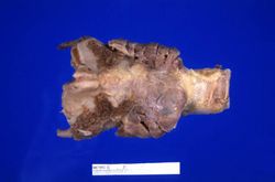
This is a gross photograph of thyroid gland taken at autopsy. The gland is only slightly enlarged and has a firm texture.
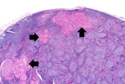
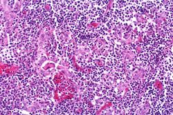
This is a low-power photomicrograph of thyroid from this case. Note the large number of blue-staining inflammatory cells in this tissue. These blue cells appear to be forming germinal centers. Some residual thyroid gland tissue can be seen in this section (arrows).
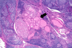
This is a higher-power photomicrograph of thyroid from this case showing the inflammatory cells and the residual thyroid tissue (arrow).
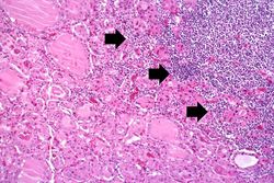
This is a high-power photomicrograph showing the inflammatory cells infiltrating into the residual thyroid tissue (arrows).
This is a high-power photomicrograph showing the lymphocytes and plasma cells surrounding the thyroid gland epithelium.
Virtual Microscopy[edit]
Study Questions[edit]
Additional Resources[edit]
Reference[edit]
- eMedicine Medical Library: Hashimoto Thyroiditis
- eMedicine Medical Library: Hypothyroidism
- Merck Manual: Overview of Thyroid Function
- Merck Manual: Hashimoto's Thyroiditis
- Merck Manual: Hypothyroidism
Journal Articles[edit]
- Agrawal P, Ogilvy-Stuart A, Lees C. Intrauterine diagnosis and management of congenital goitrous hypothyroidism. Ultrasound Obstet Gynecol 2002 May;19(5):501-5.
Images[edit]
Related IPLab Cases[edit]
Bosselated means covered with rounded protuberances.
Autoimmune disorders involve an immune response directed at the host's own cells.
A berry aneurysm is a small saccular arterial aneurysm usually found at a vessel junction in the circle of Willis. These aneurysms frequently rupture, causing a subarachnoid hemorrhage.