From Pathology Education Instructional Resource
Jump to navigation
Jump to search
Images
-
This is a gross photograph of a kidney with acute rejection from an autopsy case. Note that the kidney is swollen (edema and inflammation) and there are areas of hemorrhage throughout the kidney.
-
This is a low-power photomicrograph of the kidney that was removed from this patient. Even at this low power you can appreciate the focal accumulations of cells within this section and the diffuse cellular
infiltrate (blue dots) throughout the kidney parenchyma.
-
This is a higher-power photomicrograph demonstrating the cellular
infiltrates within this kidney section.
-
This is a higher-power photomicrograph demonstrating the cellular
infiltrates within this kidney section. Note that in addition to the diffuse cellularity, the focal accumulations of cells appear to be focused around blood vessels.
-
This is a higher-power photomicrograph demonstrating the cellular
infiltrate within the interstitium and around the small blood vessel in the center of the image.
-
This is a higher-power photomicrograph demonstrating the cellular
infiltrate within the interstitium. There is some degeneration (coagulative necrosis) of tubules and glomeruli.
-
This high-power photomicrograph demonstrates the cellular
infiltrate within the interstitium and in the wall of the blood vessel on the left.
-
This high-power photomicrograph demonstrates the cellular
infiltrate within the interstitium (1) and in the wall of the blood vessel (2).
-
This is a high-power photomicrograph of cells infiltrating the wall of the blood vessel.
-
This high-power photomicrograph demonstrates the cellular
infiltrate within the interstitium and cells within the renal tubules.
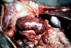 This is a gross photograph of a kidney with acute rejection from an autopsy case. Note that the kidney is swollen (edema and inflammation) and there are areas of hemorrhage throughout the kidney.
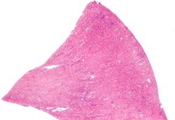 This is a low-power photomicrograph of the kidney that was removed from this patient. Even at this low power you can appreciate the focal accumulations of cells within this section and the diffuse cellular infiltrate (blue dots) throughout the kidney parenchyma.
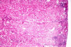 This is a higher-power photomicrograph demonstrating the cellular infiltrates within this kidney section.
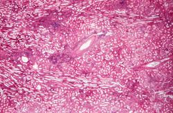 This is a higher-power photomicrograph demonstrating the cellular infiltrates within this kidney section. Note that in addition to the diffuse cellularity, the focal accumulations of cells appear to be focused around blood vessels.
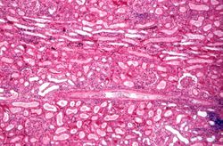 This is a higher-power photomicrograph demonstrating the cellular infiltrate within the interstitium and around the small blood vessel in the center of the image.
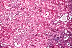 This is a higher-power photomicrograph demonstrating the cellular infiltrate within the interstitium. There is some degeneration (coagulative necrosis) of tubules and glomeruli.
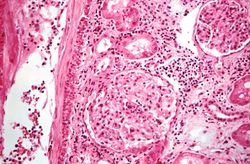 This high-power photomicrograph demonstrates the cellular infiltrate within the interstitium and in the wall of the blood vessel on the left.
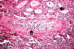 This high-power photomicrograph demonstrates the cellular infiltrate within the interstitium (1) and in the wall of the blood vessel (2).
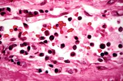 This is a high-power photomicrograph of cells infiltrating the wall of the blood vessel.
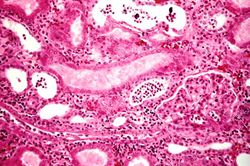 This high-power photomicrograph demonstrates the cellular infiltrate within the interstitium and cells within the renal tubules.


