From Pathology Education Instructional Resource
Jump to navigation
Jump to search
Images
-
This is a gross photograph of section of heart tissue from this case. The tissue was firm and had a waxy texture. If you use your imagination you can see pale yellow areas within this tissue which represent the amyloid deposits.
-
This is a low power photomicrograph of the heart tissue from this case. At this magnification the structure looks relatively normal.
-
This is a higher-power photomicrograph of the heart tissue from this case. Note the amyloid deposition throughout the myocardium (1) as well as deposition in the wall of the blood vessel (2).
-
This is a higher-power photomicrograph of extracellular amyloid (1) and deposition of amyloid in the vessel wall (2).
-
This is a special stain for amyloid (Luxol PAS) demonstrating the amyloid (1) and fibrosis (2) in the myocardium. The amyloid is darker purple/magenta and tends to be more amorphous. The fibrosis is pink and more fibrillar.
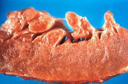 This is a gross photograph of section of heart tissue from this case. The tissue was firm and had a waxy texture. If you use your imagination you can see pale yellow areas within this tissue which represent the amyloid deposits.
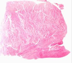 This is a low power photomicrograph of the heart tissue from this case. At this magnification the structure looks relatively normal.
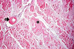 This is a higher-power photomicrograph of the heart tissue from this case. Note the amyloid deposition throughout the myocardium (1) as well as deposition in the wall of the blood vessel (2).
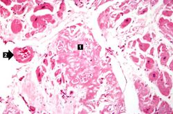 This is a higher-power photomicrograph of extracellular amyloid (1) and deposition of amyloid in the vessel wall (2).
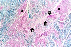 This is a special stain for amyloid (Luxol PAS) demonstrating the amyloid (1) and fibrosis (2) in the myocardium. The amyloid is darker purple/magenta and tends to be more amorphous. The fibrosis is pink and more fibrillar.


