From Pathology Education Instructional Resource
Jump to navigation
Jump to search
Images
-
This is a photograph of a section of lung with an apical lesion. This lesion represents an old healed lesion from Mycobacterium tuberculosis infection.
-
This is a low-power photomicrograph of lung tissue with multiple circumscribed nodules - granulomas (arrows).
-
This is a higher-power photomicrograph of a TB granuloma. Note the eosinophilic material in the center of this granuloma (
caseous necrosis) and the epithelioid macrophages and giant cells around the periphery.
-
This is a higher-power photomicrograph of a TB granuloma. The area of
caseous necrosis is on the left side of the image, there are multinucleated giant cells and epithelioid macrophages throughout the remainder of the tissue.
-
High-power photomicrograph of a TB granuloma with multinucleated giant cells adjacent to an area of
caseous necrosis (to the left).
-
This is a high-power (oil immersion) photomicrograph of granuloma stained with an acid-fast stain. Mycobacterium tuberculosis bacilli stain red.
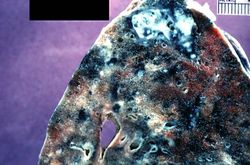 This is a photograph of a section of lung with an apical lesion. This lesion represents an old healed lesion from Mycobacterium tuberculosis infection.
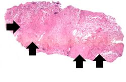 This is a low-power photomicrograph of lung tissue with multiple circumscribed nodules - granulomas (arrows).
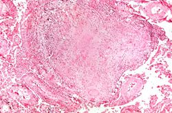 This is a higher-power photomicrograph of a TB granuloma. Note the eosinophilic material in the center of this granuloma (caseous necrosis) and the epithelioid macrophages and giant cells around the periphery.
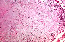 This is a higher-power photomicrograph of a TB granuloma. The area of caseous necrosis is on the left side of the image, there are multinucleated giant cells and epithelioid macrophages throughout the remainder of the tissue.
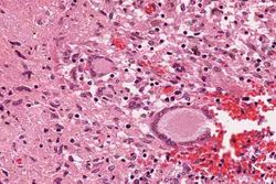 High-power photomicrograph of a TB granuloma with multinucleated giant cells adjacent to an area of caseous necrosis (to the left).
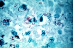 This is a high-power (oil immersion) photomicrograph of granuloma stained with an acid-fast stain. Mycobacterium tuberculosis bacilli stain red.


