From Pathology Education Instructional Resource
Jump to navigation
Jump to search
Images
-
This angiogram of the abdominal viscera demonstrates numerous aneurysms throughout the mesenteric circulation (arrows).
-
This angiogram of the liver also demonstrates numerous aneurysms throughout the hepatic circulation (arrows).
-
This angiogram of the kidneys demonstrates numerous aneurysmal dilatations in the renal circulation (arrows).
-
This is a low-power photomicrograph of a mesenteric vessel from this case of polyarteritis nodosa (arrow). The vessel is completely occluded by thrombotic material and the vessel wall is infiltrated with inflammatory cells.
-
This is a higher-power photomicrograph of this mesenteric vessel. Note the thrombotic material occluding the vessel (arrows) and the inflammatory cell
infiltrate in the wall of the vessel and in the surrounding adventitia.
-
his is another example of a mesenteric artery from this case. There is a marked inflammatory cell response surrounding this vessel, fresh hemorrhage (1), and thrombotic material (2).
-
This is a high-power photomicrograph of the vessel wall. There is hemorrhage and infiltration with inflammatory cells--primarily neutrophils (arrows).
-
This is a high-power photomicrograph of a small vessel with a rim of fibrinoid necrosis (arrow).
-
This is a low-power photomicrograph of the adrenal gland. There is an area of necrosis in the adrenal (1) and an affected vessel adjacent to the adrenal (2).
-
This is a higher-power photomicrograph of the affected vessel from the previous image. The vessel wall is infiltrated with inflammatory cells and the vessel lumen is completely occluded (arrow).
-
This is a low-power photomicrograph of the heart. There are areas of fibrosis in the myocardium (arrows). Note that the large epicardial coronary artery is normal.
-
This is a higher-power photomicrograph of the affected vessels in the heart (arrows). There are areas of fibrosis (old infarcts) in the myocardium adjacent to these affected vessels.
-
This is a high-power photomicrograph of the affected vessel in the heart. The vessel lumen is completely occluded.
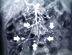 This angiogram of the abdominal viscera demonstrates numerous aneurysms throughout the mesenteric circulation (arrows).
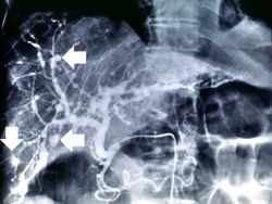 This angiogram of the liver also demonstrates numerous aneurysms throughout the hepatic circulation (arrows).
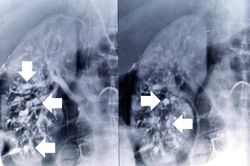 This angiogram of the kidneys demonstrates numerous aneurysmal dilatations in the renal circulation (arrows).
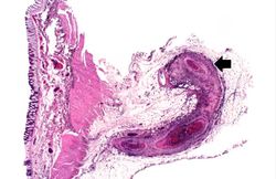 This is a low-power photomicrograph of a mesenteric vessel from this case of polyarteritis nodosa (arrow). The vessel is completely occluded by thrombotic material and the vessel wall is infiltrated with inflammatory cells.
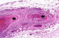 This is a higher-power photomicrograph of this mesenteric vessel. Note the thrombotic material occluding the vessel (arrows) and the inflammatory cell infiltrate in the wall of the vessel and in the surrounding adventitia.
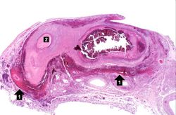 his is another example of a mesenteric artery from this case. There is a marked inflammatory cell response surrounding this vessel, fresh hemorrhage (1), and thrombotic material (2).
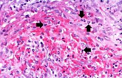 This is a high-power photomicrograph of the vessel wall. There is hemorrhage and infiltration with inflammatory cells--primarily neutrophils (arrows).
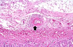 This is a high-power photomicrograph of a small vessel with a rim of fibrinoid necrosis (arrow).
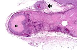 This is a low-power photomicrograph of the adrenal gland. There is an area of necrosis in the adrenal (1) and an affected vessel adjacent to the adrenal (2).
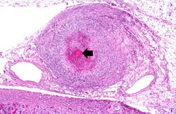 This is a higher-power photomicrograph of the affected vessel from the previous image. The vessel wall is infiltrated with inflammatory cells and the vessel lumen is completely occluded (arrow).
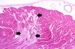 This is a low-power photomicrograph of the heart. There are areas of fibrosis in the myocardium (arrows). Note that the large epicardial coronary artery is normal.
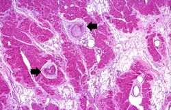 This is a higher-power photomicrograph of the affected vessels in the heart (arrows). There are areas of fibrosis (old infarcts) in the myocardium adjacent to these affected vessels.
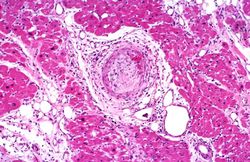 This is a high-power photomicrograph of the affected vessel in the heart. The vessel lumen is completely occluded.


