IPLab:Lab 6:Hashimoto's Thyroiditis: Difference between revisions
Jump to navigation
Jump to search
No edit summary |
No edit summary |
||
| Line 6: | Line 6: | ||
File:IPLab6Hashimoto4.jpg|This is another view of thyroid gland filled with inflammatory cells forming germinal centers (arrows). | File:IPLab6Hashimoto4.jpg|This is another view of thyroid gland filled with inflammatory cells forming germinal centers (arrows). | ||
File:IPLab6Hashimoto5.jpg|This is a higher-power photomicrograph of thyroid from this case showing the inflammatory cells and the residual thyroid tissue. | File:IPLab6Hashimoto5.jpg|This is a higher-power photomicrograph of thyroid from this case showing the inflammatory cells and the residual thyroid tissue. | ||
File:IPLab6Hashimoto6.jpg| | File:IPLab6Hashimoto6.jpg|This is another higher-power photomicrograph of thyroid from this case showing the inflammatory cells and the residual thyroid tissue. | ||
File:IPLab6Hashimoto7.jpg| | File:IPLab6Hashimoto7.jpg|This is a high-power photomicrograph showing the inflammatory cells infiltrating into the residual thyroid tissue (arrows). | ||
File:IPLab6Hashimoto8.jpg| | File:IPLab6Hashimoto8.jpg|This is a high-power photomicrograph showing the lymphocytes and plasma cells surrounding the thyroid gland epithelium. | ||
File:IPLab6Hashimoto9.jpg| | File:IPLab6Hashimoto9.jpg|This high-power photomicrograph shows more clearly the lymphocytes and plasma cells surrounding the thyroid gland epithelium. Large, eosinophilic, degenerating thyroid gland cells (Hurthle cells) can be seen in this section (arrows). | ||
</gallery> | </gallery> | ||
Revision as of 12:48, 20 August 2013
Images
-
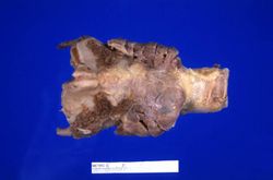 This is a gross photograph of thyroid gland taken at autopsy. The gland is only slightly enlarged and has a firm texture.
 -
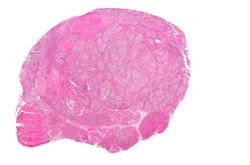 This is a low-power photomicrograph of thyroid from this case. Note that the tissue is more cellular than one would expect and there does not appear to be normal colloid-filled blue spaces in this gland.
 -
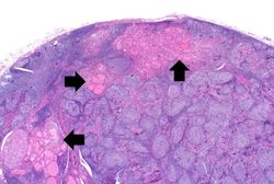 This is a higher-power photomicrograph of thyroid from this case. Note the large number of blue-staining inflammatory cells in this tissue. These cells appear to be forming germinal centers. Some residual thyroid gland tissue can be seen in this section (arrows).
 -
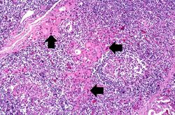
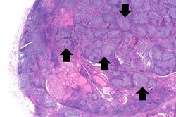 This is another view of thyroid gland filled with inflammatory cells forming germinal centers (arrows).
 -
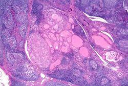 This is a higher-power photomicrograph of thyroid from this case showing the inflammatory cells and the residual thyroid tissue.
 -
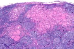 This is another higher-power photomicrograph of thyroid from this case showing the inflammatory cells and the residual thyroid tissue.
 -
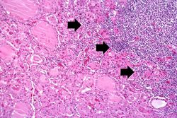 This is a high-power photomicrograph showing the inflammatory cells infiltrating into the residual thyroid tissue (arrows).
 -
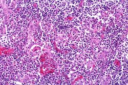 This is a high-power photomicrograph showing the lymphocytes and plasma cells surrounding the thyroid gland epithelium.
 -
 This high-power photomicrograph shows more clearly the lymphocytes and plasma cells surrounding the thyroid gland epithelium. Large, eosinophilic, degenerating thyroid gland cells (Hurthle cells) can be seen in this section (arrows).


