Difference between revisions of "IPLab:Lab 13:Cystic Fibrosis"
Seung Park (talk | contribs) (→Virtual Microscopy) |
(→Autopsy Findings) |
||
| Line 1: | Line 1: | ||
== Clinical Summary == | == Clinical Summary == | ||
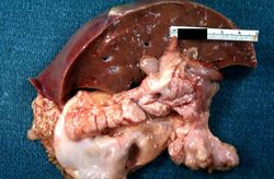
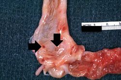
This white female infant was the product of an uncomplicated term delivery, though meconium staining was noted at birth. During the first day post-partum, the infant's abdomen became progressively distended and a meconium ileus was suspected. Surgery confirmed the presence of a meconium ileus and a section of perforated atretic jejunum proximal to the ileus was resected. Eight days later, the patient's condition had deteriorated. A second operation revealed a segment of necrotic bowel, which was removed. Subsequently the infant's pulmonary function deteriorated and she required frequent suctioning. She developed repeated episodes of pneumonia (E. coli and Pseudomonas grew out on cultures) complicated by atelectasis secondary to pneumothorax. The patient died at 25-days-of-age in respiratory failure. | This white female infant was the product of an uncomplicated term delivery, though meconium staining was noted at birth. During the first day post-partum, the infant's abdomen became progressively distended and a meconium ileus was suspected. Surgery confirmed the presence of a meconium ileus and a section of perforated atretic jejunum proximal to the ileus was resected. Eight days later, the patient's condition had deteriorated. A second operation revealed a segment of necrotic bowel, which was removed. Subsequently the infant's pulmonary function deteriorated and she required frequent suctioning. She developed repeated episodes of pneumonia (E. coli and Pseudomonas grew out on cultures) complicated by atelectasis secondary to pneumothorax. The patient died at 25-days-of-age in respiratory failure. | ||
| − | |||
| − | |||
| − | |||
== Images == | == Images == | ||
Revision as of 21:35, 9 July 2020
Contents
Clinical Summary[edit]
This white female infant was the product of an uncomplicated term delivery, though meconium staining was noted at birth. During the first day post-partum, the infant's abdomen became progressively distended and a meconium ileus was suspected. Surgery confirmed the presence of a meconium ileus and a section of perforated atretic jejunum proximal to the ileus was resected. Eight days later, the patient's condition had deteriorated. A second operation revealed a segment of necrotic bowel, which was removed. Subsequently the infant's pulmonary function deteriorated and she required frequent suctioning. She developed repeated episodes of pneumonia (E. coli and Pseudomonas grew out on cultures) complicated by atelectasis secondary to pneumothorax. The patient died at 25-days-of-age in respiratory failure.
Images[edit]
Virtual Microscopy[edit]
Pancreas[edit]
Duodenum[edit]
Study Questions[edit]
Additional Resources[edit]
Reference[edit]
- eMedicine Medical Library: Cystic Fibrosis
- eMedicine Medical Library: Cystic Fibrosis Treatment and Management
- eMedicine Medical Library: Sinonasal Manifestations of Cystic Fibrosis
- Merck Manual: Cystic Fibrosis (CF)
Journal Articles[edit]
- De Boeck K, Alifier M, Vandeputte S. Sputum induction in young cystic fibrosis patients. Eur Respir J 2000 Jul;16(1):91-4.
- Aurora P, Wade A, Whitmore P, Whitehead B. A model for predicting life expectancy of children with cystic fibrosis. Eur Respir J 2000 Dec;16(6):1056-60.
Images[edit]
| |||||
Meconium is a dark-green mucilaginous mixture of intestinal secretions and amniotic fluid which is found in the intestine of a full-term fetus. Meconium-stained amniotic fluid, found at delivery, may be an indication of perinatal asphyxia.
Meconium ileus occurs when abnormally viscid meconium completely obstructs the ileum of a newborn.
In alcoholics, aspiration pneumonia is common--bacteria enter the lung via aspiration of gastric contents.
Atelectasis is the collapse of an airway and lung, regardless of the cause, resulting in reduced or absent gas exchange.
A pneumothorax is an accumulation of gas in the pleural space.
Cirrhosis is a liver disease characterized by necrosis, fibrosis, loss of normal liver architecture, and hyperplastic nodules.
