IPLab:Lab 6:Senile Amyloidosis: Difference between revisions
Seung Park (talk | contribs) No edit summary |
|||
| Line 1: | Line 1: | ||
== Clinical Summary == | == Clinical Summary == | ||
This 87-year-old black male diabetic was admitted for amputation of the lower extremity involved by atherosclerotic gangrene and osteomyelitis. Following amputation, the patient's course was one of progressive deterioration. Laboratory studies immediately prior to death revealed a blood glucose of 840 mg/dL and a serum CO2 (bicarbonate) of 8.5 mmol/L. | This 87-year-old black male diabetic was admitted for amputation of the lower extremity involved by atherosclerotic gangrene and osteomyelitis. Following amputation, the patient's course was one of progressive deterioration. Laboratory studies immediately prior to death revealed a blood glucose of 840 mg/dL and a serum CO2 (bicarbonate) of 8.5 mmol/L. | ||
At autopsy the heart weighed 540 grams. The endocardial surface of both atria presented a mottled gray-red and gray-white appearance. Multiple sections through the myocardium revealed a marked 'pallor' of the muscle. | |||
== Autopsy Findings == | == Autopsy Findings == | ||
Revision as of 19:23, 8 July 2020
Clinical Summary
This 87-year-old black male diabetic was admitted for amputation of the lower extremity involved by atherosclerotic gangrene and osteomyelitis. Following amputation, the patient's course was one of progressive deterioration. Laboratory studies immediately prior to death revealed a blood glucose of 840 mg/dL and a serum CO2 (bicarbonate) of 8.5 mmol/L.
At autopsy the heart weighed 540 grams. The endocardial surface of both atria presented a mottled gray-red and gray-white appearance. Multiple sections through the myocardium revealed a marked 'pallor' of the muscle.
Autopsy Findings
The heart weighed 540 grams. The endocardial surface of both atria presented a mottled gray-red and gray-white appearance. Multiple sections through the myocardium revealed a marked 'pallor' of the muscle.
Images
-
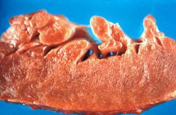 This is a gross photograph of section of heart tissue from this case. The tissue was firm and had a waxy texture. If you use your imagination you can see pale yellow areas within this tissue which represent the amyloid deposits.
 -
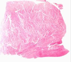 This is a low power photomicrograph of the heart tissue from this case. At this magnification the structure looks relatively normal.
 -
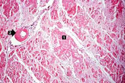 This is a higher-power photomicrograph of the heart tissue from this case. Note the amyloid deposition throughout the myocardium (1) as well as deposition in the wall of the blood vessel (2).
 -
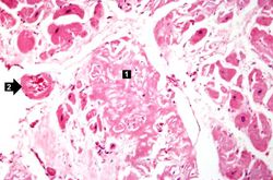 This is a higher-power photomicrograph of extracellular amyloid (1) and deposition of amyloid in the vessel wall (2).
 -
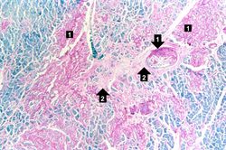 This is a special stain for amyloid (Luxol PAS) demonstrating the amyloid (1) and fibrosis (2) in the myocardium. The amyloid is darker purple/magenta and tends to be more amorphous. The fibrosis is pink and more fibrillar.


Virtual Microscopy
Study Questions
Additional Resources
Reference
- eMedicine Medical Library: Amyloidosis
- eMedicine Medical Library: Transthyretin-Related Amyloidosis
- Merck Manual: Amyloidosis
Journal Articles
- Moyssakis I, Triposkiadis F, Rallidis L, Hawkins P, Kyriakidis M, Nihoyannopoulos P. Echocardiographic features of primary, secondary and familial amyloidosis. Eur J Clin Invest 1999 Jun;29(6):484-9.