IPLab:Lab 6:Acute Rejection: Difference between revisions
Seung Park (talk | contribs) |
Seung Park (talk | contribs) No edit summary |
||
| Line 18: | Line 18: | ||
File:IPLab6AcuteRejection10.jpg|This high-power photomicrograph demonstrates the cellular infiltrate within the interstitium and cells within the renal tubules. | File:IPLab6AcuteRejection10.jpg|This high-power photomicrograph demonstrates the cellular infiltrate within the interstitium and cells within the renal tubules. | ||
</gallery> | </gallery> | ||
== Virtual Microscopy == | |||
<peir-vm>IPLab6AcuteRejection</peir-vm> | |||
== Study Questions == | == Study Questions == | ||
Revision as of 11:19, 3 January 2014
Clinical Summary
This 34-year-old white male with end-stage chronic glomerulonephritis had been receiving hemodialysis three times per week for 4 months when he was admitted to the hospital for a living related-donor transplantation from his mother. Other than the kidney disease, the patient was in good health. The transplant was performed successfully with no complications. However, eight days later, transplant rejection necessitated returning the patient to the operating room for a nephrectomy of the transplanted kidney. After the nephrectomy, the patient did quite well, was returned to hemodialysis, and was discharged home in good condition.
Autopsy Findings
The kidney weighed 240 grams and was edematous. The capsule stripped with ease to reveal a pale tan-brown cortex which was irregularly red-mottled. Upon sectioning, the cortical band was ill-defined, and the corticomedullary junction was not well-demarcated. The renal papillae were edematous and the renal pelvis displayed generalized petechial hemorrhages which extended through the 7-cm segment of ureter to a diffusely hemorrhagic terminal portion.
Images
-
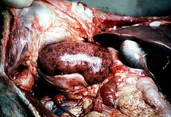 This is a gross photograph of a kidney with acute rejection from an autopsy case. Note that the kidney is swollen (edema and inflammation) and there are areas of hemorrhage throughout the kidney.
 -
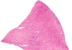 This is a low-power photomicrograph of the kidney that was removed from this patient. Even at this low power you can appreciate the focal accumulations of cells within this section and the diffuse cellular infiltrate (blue dots) throughout the kidney parenchyma.
 -
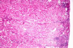 This is a higher-power photomicrograph demonstrating the cellular infiltrates within this kidney section.
 -
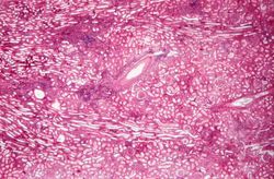 This is a higher-power photomicrograph demonstrating the cellular infiltrates within this kidney section. Note that in addition to the diffuse cellularity, the focal accumulations of cells appear to be focused around blood vessels.
 -
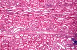 This is a higher-power photomicrograph demonstrating the cellular infiltrate within the interstitium and around the small blood vessel in the center of the image.
 -
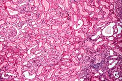 This is a higher-power photomicrograph demonstrating the cellular infiltrate within the interstitium. There is some degeneration (coagulative necrosis) of tubules and glomeruli.
 -
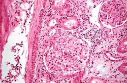 This high-power photomicrograph demonstrates the cellular infiltrate within the interstitium and in the wall of the blood vessel on the left.
 -
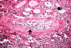 This high-power photomicrograph demonstrates the cellular infiltrate within the interstitium (1) and in the wall of the blood vessel (2).
 -
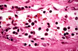 This is a high-power photomicrograph of cells infiltrating the wall of the blood vessel.
 -
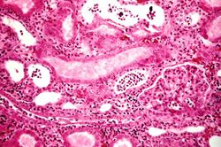 This high-power photomicrograph demonstrates the cellular infiltrate within the interstitium and cells within the renal tubules.


Virtual Microscopy
Study Questions
Additional Resources
Reference
- eMedicine Medical Library: Assessment and Management of the Renal Transplant Patient
- eMedicine Medical Library: Renal Transplantation
- Merck Manual: Nephrotic Syndrome
- Merck Manual: Chronic Kidney Disease
- Merck Manual: Hemodialysis
- Merck Manual: Kidney Transplantation
Journal Articles
- Matas AJ. Impact of acute rejection on development of chronic rejection in pediatric renal transplant recipients. Pediatr Transplant 2000 May;4(2):92-9.
Images
Related IPLab Cases
- Lab 1: Kidney: Infarction (Coagulative Necrosis)
- Lab 6: Kidney: Chronic Transplant Rejection
- Lab 10: Kidney: Candidiasis