IPLab:Lab 6:Tuberculosis: Difference between revisions
No edit summary |
Seung Park (talk | contribs) No edit summary |
||
| Line 1: | Line 1: | ||
== Clinical Summary == | |||
This 29-year-old black female had a history of scleroderma involving the lung, kidney, heart, and skin. Her main clinical problems centered on her restrictive lung disease. She was able to live at home with supplemental oxygen but recently she had developed edema, chest pain, weakness, light-headedness, and a loss of appetite. The patient was admitted to the hospital with a working diagnosis of congestive heart failure brought on by her lung disease. Echocardiographic evaluation revealed a pericardial effusion that was tapped. Soon after this procedure her respiratory status degenerated and she required intubation. Despite aggressive supportive treatment for her cardiac and pulmonary problems, she could not be weaned from the ventilator. Two weeks after admission she became febrile and Gram positive cocci were isolated from sputum culture. She was placed on antibiotics but her condition deteriorated and she developed bradycardia followed by electromechanical dissociation (EMD). | |||
== Autopsy Findings == | |||
Upon opening the thorax there was 600 cc of cloudy serous fluid in each hemithorax and 100 cc of similar fluid in the pericardial sac. The heart weighed 530 grams and there was thickening of both the left and right ventricular walls. The liver weighed 1880 grams and was congested. The spleen weighed 200 grams and was also congested. The combined lung weight was 1875 grams; the lungs were markedly fibrotic with severe emphysema. In addition, dermal thickening was evident throughout the body and the wall of the esophagus was thickened and firm. | |||
== Images == | == Images == | ||
<gallery heights="250px" widths="250px"> | <gallery heights="250px" widths="250px"> | ||
Revision as of 14:25, 21 August 2013
Clinical Summary
This 29-year-old black female had a history of scleroderma involving the lung, kidney, heart, and skin. Her main clinical problems centered on her restrictive lung disease. She was able to live at home with supplemental oxygen but recently she had developed edema, chest pain, weakness, light-headedness, and a loss of appetite. The patient was admitted to the hospital with a working diagnosis of congestive heart failure brought on by her lung disease. Echocardiographic evaluation revealed a pericardial effusion that was tapped. Soon after this procedure her respiratory status degenerated and she required intubation. Despite aggressive supportive treatment for her cardiac and pulmonary problems, she could not be weaned from the ventilator. Two weeks after admission she became febrile and Gram positive cocci were isolated from sputum culture. She was placed on antibiotics but her condition deteriorated and she developed bradycardia followed by electromechanical dissociation (EMD).
Autopsy Findings
Upon opening the thorax there was 600 cc of cloudy serous fluid in each hemithorax and 100 cc of similar fluid in the pericardial sac. The heart weighed 530 grams and there was thickening of both the left and right ventricular walls. The liver weighed 1880 grams and was congested. The spleen weighed 200 grams and was also congested. The combined lung weight was 1875 grams; the lungs were markedly fibrotic with severe emphysema. In addition, dermal thickening was evident throughout the body and the wall of the esophagus was thickened and firm.
Images
-
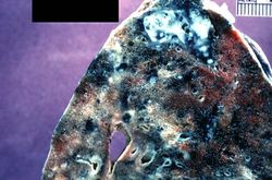 This is a photograph of a section of lung with an apical lesion. This lesion represents an old healed lesion from Mycobacterium tuberculosis infection.
 -
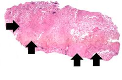 This is a low-power photomicrograph of lung tissue with multiple circumscribed nodules - granulomas (arrows).
 -
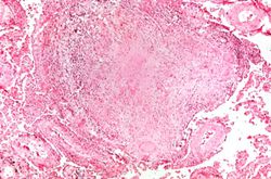 This is a higher-power photomicrograph of a TB granuloma. Note the eosinophilic material in the center of this granuloma (caseous necrosis) and the epithelioid macrophages and giant cells around the periphery.
 -
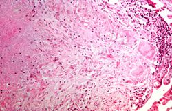 This is a higher-power photomicrograph of a TB granuloma. The area of caseous necrosis is on the left side of the image, there are multinucleated giant cells and epithelioid macrophages throughout the remainder of the tissue.
 -
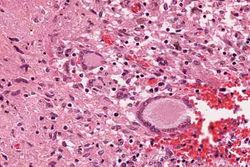 High-power photomicrograph of a TB granuloma with multinucleated giant cells adjacent to an area of caseous necrosis (to the left).
 -
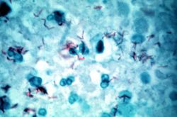 This is a high-power (oil immersion) photomicrograph of granuloma stained with an acid-fast stain. Mycobacterium tuberculosis bacilli stain red.


