IPLab:Lab 6:Tuberculosis: Difference between revisions
Jump to navigation
Jump to search
Created page with "== Images == <gallery heights="250px" widths="250px"> File:IPLab6TB1.jpg| File:IPLab6TB2.jpg| File:IPLab6TB3.jpg| File:IPLab6TB4.jpg| File:IPLab6TB5.jpg| </gallery> {{IPLab 6..." |
No edit summary |
||
| Line 1: | Line 1: | ||
== Images == | == Images == | ||
<gallery heights="250px" widths="250px"> | <gallery heights="250px" widths="250px"> | ||
File:IPLab6TB1.jpg| | File:IPLab6TB1.jpg|This is a photograph of a section of lung with an apical lesion. This lesion represents an old healed lesion from Mycobacterium tuberculosis infection. | ||
File:IPLab6TB2.jpg| | File:IPLab6TB2.jpg|This is a low-power photomicrograph of lung tissue with multiple circumscribed nodules - granulomas (arrows). | ||
File:IPLab6TB3.jpg| | File:IPLab6TB3.jpg|This is a higher-power photomicrograph of a TB granuloma. Note the eosinophilic material in the center of this granuloma (caseous necrosis) and the epithelioid macrophages and giant cells around the periphery. | ||
File:IPLab6TB4.jpg| | File:IPLab6TB4.jpg|This is a higher-power photomicrograph of a TB granuloma. The area of caseous necrosis is on the left side of the image, there are multinucleated giant cells and epithelioid macrophages throughout the remainder of the tissue. | ||
File:IPLab6TB5.jpg| | File:IPLab6TB5.jpg|High-power photomicrograph of a TB granuloma with multinucleated giant cells adjacent to an area of caseous necrosis (to the left). | ||
File:IPLab6TB6.jpg|This is a high-power (oil immersion) photomicrograph of granuloma stained with an acid-fast stain. Mycobacterium tuberculosis bacilli stain red. | |||
</gallery> | </gallery> | ||
Revision as of 15:14, 20 August 2013
Images
-
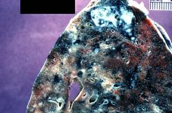 This is a photograph of a section of lung with an apical lesion. This lesion represents an old healed lesion from Mycobacterium tuberculosis infection.
 -
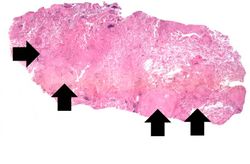 This is a low-power photomicrograph of lung tissue with multiple circumscribed nodules - granulomas (arrows).
 -
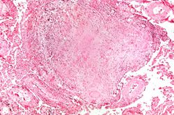 This is a higher-power photomicrograph of a TB granuloma. Note the eosinophilic material in the center of this granuloma (caseous necrosis) and the epithelioid macrophages and giant cells around the periphery.
 -
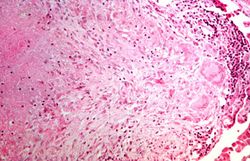 This is a higher-power photomicrograph of a TB granuloma. The area of caseous necrosis is on the left side of the image, there are multinucleated giant cells and epithelioid macrophages throughout the remainder of the tissue.
 -
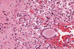 High-power photomicrograph of a TB granuloma with multinucleated giant cells adjacent to an area of caseous necrosis (to the left).
 -
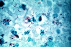 This is a high-power (oil immersion) photomicrograph of granuloma stained with an acid-fast stain. Mycobacterium tuberculosis bacilli stain red.


