IPLab:Lab 6:Amyloidosis: Difference between revisions
Jump to navigation
Jump to search
Seung Park (talk | contribs) |
Seung Park (talk | contribs) No edit summary |
||
| Line 19: | Line 19: | ||
File:IPLab6Amyloid11.jpg|This photomicrograph of the tongue demonstrates extensive amyloid deposits (1) separating the skeletal muscle fibers of the tongue. In many cases the amyloid encircles the muscle fibers (2) and these muscle fibers are atrophied. | File:IPLab6Amyloid11.jpg|This photomicrograph of the tongue demonstrates extensive amyloid deposits (1) separating the skeletal muscle fibers of the tongue. In many cases the amyloid encircles the muscle fibers (2) and these muscle fibers are atrophied. | ||
</gallery> | </gallery> | ||
== Virtual Microscopy == | |||
<peir-vm>IPLab6Amyloid</peir-vm> | |||
== Study Questions == | == Study Questions == | ||
Revision as of 11:20, 3 January 2014
Clinical Summary
This 46-year-old white male with a long-standing history of rheumatoid arthritis was admitted for treatment of pneumonia. Subsequently, complications associated with lung abscesses, empyema, and septicemia led to the patient's death.
Autopsy Findings
The liver weighed 2600 grams. It was yellowish-tan in color and cut with difficulty (fibrosis?). No other pathological changes were noted except for pneumonia and lung abscesses.
Images
-
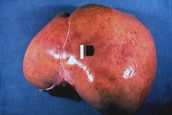 This is a gross picture of liver from this case. Note the pale, swollen appearance of this liver.
 -
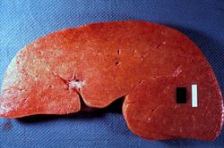 This is a gross picture of the cut surface of the liver from this case. The liver tissue is firm and has a waxy appearance--although this is difficult to appreciate in an image.
 -
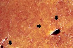 This is a closer view of the cut surface of this liver. The pale waxy material can be seen within the hepatic tissue (arrows).
 -
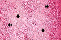 This is a low-power photomicrograph of liver tissue from this case. Note the eosinophilic hyaline material (1) present within and between the hepatic tissue (2). There is marked distortion of lobular architecture by the amyloid.
 -
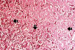 This is a higher-power photomicrograph showing the amyloid deposits (1) between hepatocytes (2).
 -
 This is a low-power photomicrograph of liver tissue stained with Congo red (orange color in slide). Congo red reacts with amyloid and gives it an orange color (arrows).
 -
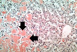 This is a high-power view of liver tissue stained with Congo red. The orange amyloid material (arrows) is seen clearly between liver parenchymal cells.
 -
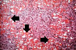 This is a photomicrograph of Congo-red-stained liver tissue viewed with partially polarized light. Although not well demonstrated in this image, Congo-red-stained amyloid viewed through polarized light should give off a classic “apple green” birefringence (arrows).
 -
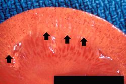 This is a gross photograph of kidney from this case. Note the pale yellow material within the cortex (arrows). This is indicative of amyloid within the cortex and the glomeruli. Also note that there are multiple red spots in the cortex. These represent congested glomeruli due to the vascular compromise produced by the amyloid.
 -
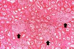 This photomicrograph of kidney demonstrates the amyloid deposits (arrows) within glomeruli.
 -
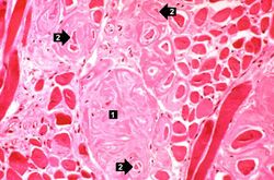 This photomicrograph of the tongue demonstrates extensive amyloid deposits (1) separating the skeletal muscle fibers of the tongue. In many cases the amyloid encircles the muscle fibers (2) and these muscle fibers are atrophied.


Virtual Microscopy
Study Questions
Additional Resources
Reference
- eMedicine Medical Library: Amyloidosis
- eMedicine Medical Library: Amyloidosis, AA (Inflammatory)
- Merck Manual: Amyloidosis
Journal Articles
- Obici L, Merlini G. AA amyloidosis: basic knowledge, unmet needs and future treatments. Swiss Med Wkly 2012 May 31;142:w13580.
Images
Related IPLab Cases