IPLab:Lab 6:Hashimoto's Thyroiditis: Difference between revisions
No edit summary |
Seung Park (talk | contribs) |
||
| Line 35: | Line 35: | ||
=== Images === | === Images === | ||
* [ | * [{{SERVER}}/library/index.php?/tags/15-endocrine/140-thyroid/141-lymphocytic_thyroiditis PEIR Digital Library: Lymphocytic Thyroiditis Images] | ||
* [http://library.med.utah.edu/WebPath/ENDOHTML/ENDOIDX.html WebPath: Endocrine Pathology] | * [http://library.med.utah.edu/WebPath/ENDOHTML/ENDOIDX.html WebPath: Endocrine Pathology] | ||
Revision as of 20:35, 29 August 2013
Clinical Summary
This was a 49-year-old woman who complained during her yearly physical examination of tiredness and difficulty concentrating. She attributed these symptoms to stress at work. She had gained weight over the last year and despite warm weather, she felt chilled without a sweater. Family history was significant for hypothyroidism in her mother and older sister. On physical examination she had a pulse of 58 bpm and a blood pressure of 138/88 mm Hg. Examination of her neck disclosed a small thyroid gland with a palpable pyramidal lobe and a firm, bosselated texture. Serum taken at this time demonstrated a total T4 of 7.0 mcg/dL and a TSH of 22.0 mcIU/ml. In addition, antithyroglobulin antibodies were positive at 1 to 640 and antimicrosomal antibodies were positive at 1 to 5120. These results supported the clinical impression of hypothyroidism; also, the texture of her thyroid gland and a positive family history suggested an autoimmune etiological factor. She was referred to an endocrinologist for further evaluation; however, before beginning treatment she died suddenly from a ruptured berry aneurysm of the middle cerebral artery.
Autopsy Findings
At autopsy, significant subarachnoid hemorrhage from the ruptured berry aneurysm was documented. In addition, the thyroid gland was mildly enlarged and firm. On cut section the tissue was slightly pale.
Images
-
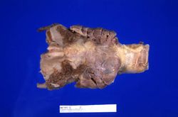 This is a gross photograph of thyroid gland taken at autopsy. The gland is only slightly enlarged and has a firm texture.
 -
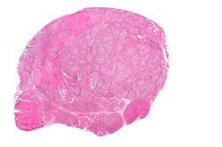 This is a low-power photomicrograph of thyroid from this case. Note that the tissue is more cellular than one would expect and there does not appear to be normal colloid-filled blue spaces in this gland.
 -
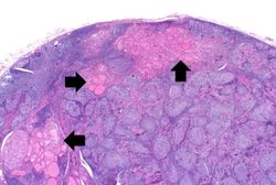 This is a higher-power photomicrograph of thyroid from this case. Note the large number of blue-staining inflammatory cells in this tissue. These cells appear to be forming germinal centers. Some residual thyroid gland tissue can be seen in this section (arrows).
 -
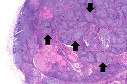 This is another view of thyroid gland filled with inflammatory cells forming germinal centers (arrows).
 -
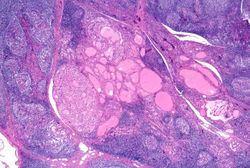 This is a higher-power photomicrograph of thyroid from this case showing the inflammatory cells and the residual thyroid tissue.
 -
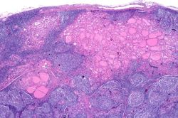 This is another higher-power photomicrograph of thyroid from this case showing the inflammatory cells and the residual thyroid tissue.
 -
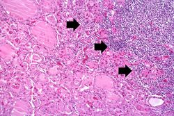 This is a high-power photomicrograph showing the inflammatory cells infiltrating into the residual thyroid tissue (arrows).
 -
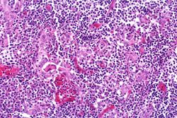 This is a high-power photomicrograph showing the lymphocytes and plasma cells surrounding the thyroid gland epithelium.
 -
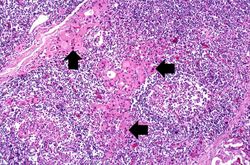 This high-power photomicrograph shows more clearly the lymphocytes and plasma cells surrounding the thyroid gland epithelium. Large, eosinophilic, degenerating thyroid gland cells (Hurthle cells) can be seen in this section (arrows).


Study Questions
Additional Resources
Reference
- eMedicine Medical Library: Hashimoto Thyroiditis
- eMedicine Medical Library: Hypothyroidism
- Merck Manual: Overview of Thyroid Function
- Merck Manual: Hashimoto's Thyroiditis
- Merck Manual: Hypothyroidism
Journal Articles
- Agrawal P, Ogilvy-Stuart A, Lees C. Intrauterine diagnosis and management of congenital goitrous hypothyroidism. Ultrasound Obstet Gynecol 2002 May;19(5):501-5.