IPLab:Lab 6:Multiple Myeloma: Difference between revisions
No edit summary |
|||
| (10 intermediate revisions by 2 users not shown) | |||
| Line 2: | Line 2: | ||
This 63-year-old female presented with the complaint of left chest pain of approximately 4 months duration. Physical examination revealed that the pain was along the distribution of the left sixth intercostal nerve. Chest film showed a posterior mediastinal mass with partial collapse of T6. A lytic lesion of the right distal clavicle was noted on subsequent radiological examination. A bone scan revealed increased uptake in thoracic vertebrae. Serum alkaline phosphatase was elevated slightly (143 U/L). Serum protein electrophoresis was normal, while urine protein electrophoresis showed a monoclonal spike in the Gamma region. A bone marrow study was non-diagnostic. | This 63-year-old female presented with the complaint of left chest pain of approximately 4 months duration. Physical examination revealed that the pain was along the distribution of the left sixth intercostal nerve. Chest film showed a posterior mediastinal mass with partial collapse of T6. A lytic lesion of the right distal clavicle was noted on subsequent radiological examination. A bone scan revealed increased uptake in thoracic vertebrae. Serum alkaline phosphatase was elevated slightly (143 U/L). Serum protein electrophoresis was normal, while urine protein electrophoresis showed a monoclonal spike in the Gamma region. A bone marrow study was non-diagnostic. | ||
A thoracotomy was performed after the unsuccessful needle biopsy. At thoracotomy, a 3-cm posterior mediastinal mass was identified that extended to within 1-2 mm of the aorta and into the interspace between the ribs. | |||
A thoracotomy was performed after | |||
== Images == | == Images == | ||
| Line 12: | Line 11: | ||
File:IPLab6MM4.jpg|This is a photograph of the vertebral column from this patient at autopsy. Notice the collapsed vertebra (1). There are multiple variably-sized white nodules (2) within the bone marrow. These are accumulations of malignant plasma cells in this case of multiple myeloma. | File:IPLab6MM4.jpg|This is a photograph of the vertebral column from this patient at autopsy. Notice the collapsed vertebra (1). There are multiple variably-sized white nodules (2) within the bone marrow. These are accumulations of malignant plasma cells in this case of multiple myeloma. | ||
</gallery> | </gallery> | ||
== Virtual Microscopy == | |||
<peir-vm>IPLab6MM</peir-vm> | |||
== Study Questions == | == Study Questions == | ||
| Line 17: | Line 19: | ||
* <spoiler text="What is the chemical nature of this type of amyloid?">Immunoglobulin light chains, usually lambda.</spoiler> | * <spoiler text="What is the chemical nature of this type of amyloid?">Immunoglobulin light chains, usually lambda.</spoiler> | ||
* <spoiler text="What is the usual organ distribution of amyloid in this class of amyloidosis?">Heart, gastrointestinal tract, peripheral nerves, skin, and tongue.</spoiler> | * <spoiler text="What is the usual organ distribution of amyloid in this class of amyloidosis?">Heart, gastrointestinal tract, peripheral nerves, skin, and tongue.</spoiler> | ||
== Additional Resources == | |||
=== Reference === | |||
* [http://emedicine.medscape.com/article/204369-overview eMedicine Medical Library: Multiple Myeloma] | |||
* [http://www.merckmanuals.com/professional/hematology_and_oncology/plasma_cell_disorders/multiple_myeloma.html Merck Manual: Multiple Myeloma] | |||
=== Journal Articles === | |||
* Rodon P, Linassier C, Gauvain JB, Benboubker L, Goupille P, Maigre M, Luthier F, Dugay J, Lucas V, Colombat P. [http://www.ncbi.nlm.nih.gov/pubmed/11168502 Multiple myeloma in elderly patients: presenting features and outcome]. ''Eur J Haematol'' 2001 Jan;66(1):11-7. | |||
=== Images === | |||
* [{{SERVER}}/library/index.php?/tags/327-multiple_myeloma PEIR Digital Library: Multiple Myeloma Images] | |||
* [{{SERVER}}/library/index.php?/tags/65-amyloidosis PEIR Digital Library: Amyloidosis Images] | |||
* [http://library.med.utah.edu/WebPath/HEMEHTML/HEMEIDX.html#6 WebPath: Myeloma] | |||
== Related IPLab Cases == | |||
* [[IPLab:Lab 6:Amyloidosis|Lab 6: Liver: Amyloidosis]] | |||
* [[IPLab:Lab 6:Senile Amyloidosis|Lab 6: Heart: Senile Amyloidosis]] | |||
{{IPLab 6}} | {{IPLab 6}} | ||
[[Category: IPLab:Lab 6]] | [[Category: IPLab:Lab 6]] | ||
Latest revision as of 19:11, 8 July 2020
Clinical Summary
This 63-year-old female presented with the complaint of left chest pain of approximately 4 months duration. Physical examination revealed that the pain was along the distribution of the left sixth intercostal nerve. Chest film showed a posterior mediastinal mass with partial collapse of T6. A lytic lesion of the right distal clavicle was noted on subsequent radiological examination. A bone scan revealed increased uptake in thoracic vertebrae. Serum alkaline phosphatase was elevated slightly (143 U/L). Serum protein electrophoresis was normal, while urine protein electrophoresis showed a monoclonal spike in the Gamma region. A bone marrow study was non-diagnostic.
A thoracotomy was performed after the unsuccessful needle biopsy. At thoracotomy, a 3-cm posterior mediastinal mass was identified that extended to within 1-2 mm of the aorta and into the interspace between the ribs.
Images
-
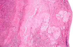 This is a low-power photomicrograph of the mediastinal mass. The mass is encapsulated and contains cellular areas (blue) and areas of pale red material.
 -
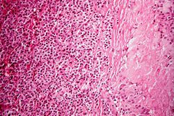 This higher-power photomicrograph shows the junction between an amorphous hyaline-appearing area (amyloid) on the right and cellular areas (plasmacytoid cells) on the left.
 -
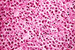 This high-power photomicrograph demonstrates the cells that make up this tissue. These cells resemble plasma cells and are the malignant cell of multiple myeloma.
 -
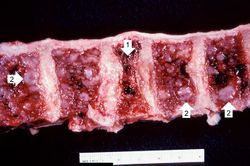 This is a photograph of the vertebral column from this patient at autopsy. Notice the collapsed vertebra (1). There are multiple variably-sized white nodules (2) within the bone marrow. These are accumulations of malignant plasma cells in this case of multiple myeloma.


Virtual Microscopy
Study Questions
Additional Resources
Reference
Journal Articles
- Rodon P, Linassier C, Gauvain JB, Benboubker L, Goupille P, Maigre M, Luthier F, Dugay J, Lucas V, Colombat P. Multiple myeloma in elderly patients: presenting features and outcome. Eur J Haematol 2001 Jan;66(1):11-7.
Images
- PEIR Digital Library: Multiple Myeloma Images
- PEIR Digital Library: Amyloidosis Images
- WebPath: Myeloma