Difference between revisions of "IPLab:Lab 6:Scleroderma"
From Pathology Education Instructional Resource
Revision as of 19:59, 20 August 2013
Images
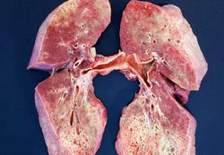
This is a gross photograph of cut section of the lungs from this patient. Note the extensive fibrosis of the lung parenchyma.
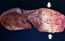
This is a gross photograph of a cut section of one lung from this patient. Note the extensive fibrosis lower lobe (arrows).
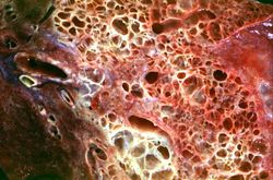
This is a closer view of the cut section of lung from this patient. Note the extensive fibrosis and the severe emphysematous changes.
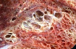
This is a closer view of the cut section of lung from this patient showing the extensive fibrosis and the severe emphysematous change.
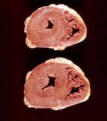
This is a gross photograph of the heart from this case. There is thickening of the left ventricular wall and some thickening of the right ventricle as well.
