Difference between revisions of "IPLab:Lab 2:Fatty Change and Cirrhosis"
(Created page with "== Images == <gallery heights="250px" widths="250px"> File:IPLab2FattyChange1.jpg|This gross photograph of liver tissue illustrates the yellowish color of the liver parenchyma...") |
|||
| Line 7: | Line 7: | ||
File:IPLab2FattyChange5.jpg|This higher-power photomicrograph of the centrilobular area gives the appearance of fatty tissue, as indicated by many empty spaces. Very few normal liver cells can be seen in this slide. A few more normal-appearing hepatocytes are present at the left portion of the slide (arrows). | File:IPLab2FattyChange5.jpg|This higher-power photomicrograph of the centrilobular area gives the appearance of fatty tissue, as indicated by many empty spaces. Very few normal liver cells can be seen in this slide. A few more normal-appearing hepatocytes are present at the left portion of the slide (arrows). | ||
File:IPLab2FattyChange6.jpg|Another view at the same power illustrates the proliferation of bile ducts in the interlobular and perichordal regions (arrows). | File:IPLab2FattyChange6.jpg|Another view at the same power illustrates the proliferation of bile ducts in the interlobular and perichordal regions (arrows). | ||
| − | File:IPLab2FattyChange7.jpg|A high-power photomicrograph of the liver parenchyma shows that each individual liver cell is filled with a large, clear droplet which represents the space remaining after lipid was dissolved by the dehydration procedure used to embed the tissue. | + | File:IPLab2FattyChange7.jpg|A high-power photomicrograph of the liver parenchyma shows that each individual liver cell is filled with a large, clear droplet which represents the space remaining after lipid was dissolved by the dehydration procedure used to embed the tissue. Note that each empty space is surrounded by a thin rim of eosinophilic cytoplasm; in many instances, the hepatocyte nucleus can be seen as well. The red body (arrow) seen within a cell in the center of the slide is an acidophilic body associated with alcoholic hepatitis. |
File:IPLab2FattyChange8.jpg|An oil red O stain for fat was performed on a frozen section of this liver tissue. The red droplets represent fat in the tissue which is typical of fatty degeneration in the liver. By using frozen sections the tissues do not have to be dehydrated through alcohol solutions and thus the fat does not get washed out. | File:IPLab2FattyChange8.jpg|An oil red O stain for fat was performed on a frozen section of this liver tissue. The red droplets represent fat in the tissue which is typical of fatty degeneration in the liver. By using frozen sections the tissues do not have to be dehydrated through alcohol solutions and thus the fat does not get washed out. | ||
File:IPLab2FattyChange9.jpg|This photomicrograph of the liver is from another patient with a history of alcohol use. There are some clear vacuoles indicating fat droplets (1) and there are numerous red-staining granular deposits within the cytoplasm of hepatocytes (2)--this is alcoholic hyalin. Alcoholic hyalin is easily distinguished from red blood cells (3) that are also present in this section. | File:IPLab2FattyChange9.jpg|This photomicrograph of the liver is from another patient with a history of alcohol use. There are some clear vacuoles indicating fat droplets (1) and there are numerous red-staining granular deposits within the cytoplasm of hepatocytes (2)--this is alcoholic hyalin. Alcoholic hyalin is easily distinguished from red blood cells (3) that are also present in this section. | ||
Revision as of 17:06, 19 August 2013
Images[edit]
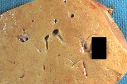
This gross photograph of liver tissue illustrates the yellowish color of the liver parenchyma. The yellow color indicates high fat content in this tissue. Compare this with the normal dark red color of liver.
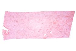
This low-power photomicrograph of liver illustrates a very pale-staining section with a uniform appearance throughout the section.
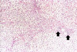
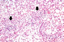
Another low-power photomicrograph illustrates again the pale, washed-out appearance of this tissue. Notice the numerous holes throughout the tissue. There are accumulations of inflammatory cells (arrows) around portal tracts.

A higher-power photomicrograph illustrates more clearly the inflammatory cells (arrows) around the portal areas.
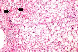
This higher-power photomicrograph of the centrilobular area gives the appearance of fatty tissue, as indicated by many empty spaces. Very few normal liver cells can be seen in this slide. A few more normal-appearing hepatocytes are present at the left portion of the slide (arrows).
Another view at the same power illustrates the proliferation of bile ducts in the interlobular and perichordal regions (arrows).
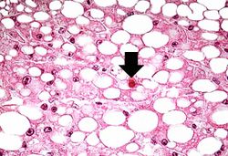
A high-power photomicrograph of the liver parenchyma shows that each individual liver cell is filled with a large, clear droplet which represents the space remaining after lipid was dissolved by the dehydration procedure used to embed the tissue. Note that each empty space is surrounded by a thin rim of eosinophilic cytoplasm; in many instances, the hepatocyte nucleus can be seen as well. The red body (arrow) seen within a cell in the center of the slide is an acidophilic body associated with alcoholic hepatitis.
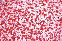
An oil red O stain for fat was performed on a frozen section of this liver tissue. The red droplets represent fat in the tissue which is typical of fatty degeneration in the liver. By using frozen sections the tissues do not have to be dehydrated through alcohol solutions and thus the fat does not get washed out.
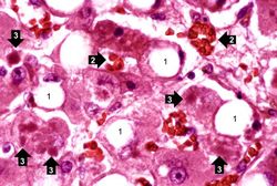
This photomicrograph of the liver is from another patient with a history of alcohol use. There are some clear vacuoles indicating fat droplets (1) and there are numerous red-staining granular deposits within the cytoplasm of hepatocytes (2)--this is alcoholic hyalin. Alcoholic hyalin is easily distinguished from red blood cells (3) that are also present in this section.
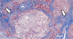
This is a low-power photomicrograph of liver stained with a trichrome stain. In this section, connective tissue stains green (arrows) and hepatic parenchymal cells are red. Note that many of the parenchymal cells have clear spaces indicating fatty degeneration. The proliferation of scar tissue between the liver lobules is the result of cirrhosis.
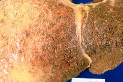
This gross photograph of liver demonstrates severe nodular cirrhosis. Note the extensive scarring of the capsule and the nodular projections of tissue through the uncut capsule in this tissue. The green color is due to the accumulation of bile pigment.
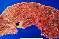
This is a cut surface of the same tissue seen in the previous slide. Note the marked nodular pattern. The paler-staining areas between the round nodules represent fibrous connective tissue.


| |||||
Cirrhosis is a liver disease characterized by necrosis, fibrosis, loss of normal liver architecture, and hyperplastic nodules.
Nodular hyperplasia of the prostate--characterized by large discrete prostatic nodules--is a common disorder in men over 50 years of age. The nodules cause the prostate to be enlarged and to have an increased weight. The human prostate is surrounded by a restrictive capsule. These nodules cause increased pressure within the capsule which leads to constriction of the urethra as it passes through the prostate. Urethral constriction leads to retention of urine.