Difference between revisions of "IPLab:Lab 2:Atrophy"
(Created page with "== Images == <gallery heights="250px" widths="250px"> File:IPLab2Atrophy1.jpg|This is a gross photograph of an atrophied testis (arrows). File:IPLab2Atrophy2.jpg| File:IPLab2A...") |
|||
| Line 2: | Line 2: | ||
<gallery heights="250px" widths="250px"> | <gallery heights="250px" widths="250px"> | ||
File:IPLab2Atrophy1.jpg|This is a gross photograph of an atrophied testis (arrows). | File:IPLab2Atrophy1.jpg|This is a gross photograph of an atrophied testis (arrows). | ||
| − | File:IPLab2Atrophy2.jpg| | + | File:IPLab2Atrophy2.jpg|This is a low-power photomicrograph of an atrophic testis. Attached to the testis are several vessels (arrow) which are part of the epididymis and the vas deferens. |
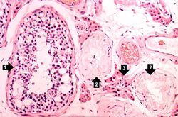
| − | File:IPLab2Atrophy3.jpg| | + | File:IPLab2Atrophy3.jpg|This is a higher-power photomicrograph of an atrophic testis. In this section there are seminiferous tubules with viable cells (1) although there are no visible spermatocytes. Other seminiferous tubules are completely acellular and have a pale pink hyaline appearance (2). |
| − | File:IPLab2Atrophy4.jpg| | + | File:IPLab2Atrophy4.jpg|This is a higher-power photomicrograph indicating loss of testicular parenchymal tissue. There are very few recognizable spermatic cells in this tissue. The cluster of cells in the upper right is a focus of interstitial or Leydig cells (arrow). These cells are not affected by the hormone-induced atrophy process. |
| − | File:IPLab2Atrophy5.jpg| | + | File:IPLab2Atrophy5.jpg|A seminiferous tubule is shown on the left containing remnants of spermatocytes (1). There are no mature sperm present. On the right-hand portion of the slide are remnants of other spermatic tubules which have completely atrophied and lost all of their spermatogenic cells (2). Note the eosinophilic Leydig cells present between the atrophic tubules (3). |
| − | File:IPLab2Atrophy6.jpg| | + | File:IPLab2Atrophy6.jpg|In this slide, the atrophy of the tubules is extending to include the Rete testes (arrow) as well. |
| − | File:IPLab2Atrophy7.jpg| | + | File:IPLab2Atrophy7.jpg|In this gross photograph of kidneys and the abdominal aorta, there is narrowing of the left renal artery at its ostium from the aorta. This atherosclerotic narrowing of the renal artery causes reduced blood pressure in the kidney whose artery is affected. The result is a characteristic Goldblatt’s hypertension syndrome. Note that the kidney with the narrowed renal artery is atrophied compared to the kidney with the normal vessel on the right. What is the cause of the atrophy in this kidney? |
| − | File:IPLab2Atrophy8.jpg| | + | File:IPLab2Atrophy8.jpg|These kidneys were removed from a patient who had blockage of one ureter leading to increased pressure in the renal pelvis. The increased pressure produced hydronephrosis (arrow) in one kidney. What is the cause of atrophy in this case? |
| − | File:IPLab2Atrophy9.jpg| | + | File:IPLab2Atrophy9.jpg|The two kidneys in this slide are from the same patient. One kidney (1) is relatively normal, although increased in size due to compensatory hypertrophy. The other kidney (2) is very small with only rudimentary nodules of renal parenchyma. This kidney had never developed and therefore this process represents hypoplasia. How does one differentiate between atrophy and hypoplasia? |
| − | File:IPLab2Atrophy10.jpg| | + | File:IPLab2Atrophy10.jpg|This gross photograph shows a normal brain (left) and a brain from a geriatric patient (right). Note the decreased size, the narrowed gyri, and the widened sulci of the brain from this octogenarian. What is the cause of atrophy in this case? |
</gallery> | </gallery> | ||
Revision as of 16:12, 19 August 2013
Images[edit]
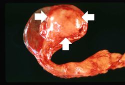
This is a gross photograph of an atrophied testis (arrows).
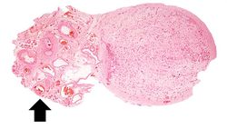
This is a low-power photomicrograph of an atrophic testis. Attached to the testis are several vessels (arrow) which are part of the epididymis and the vas deferens.
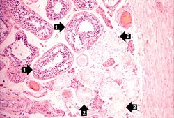
This is a higher-power photomicrograph of an atrophic testis. In this section there are seminiferous tubules with viable cells (1) although there are no visible spermatocytes. Other seminiferous tubules are completely acellular and have a pale pink hyaline appearance (2).
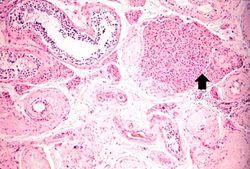
This is a higher-power photomicrograph indicating loss of testicular parenchymal tissue. There are very few recognizable spermatic cells in this tissue. The cluster of cells in the upper right is a focus of interstitial or Leydig cells (arrow). These cells are not affected by the hormone-induced atrophy process.
A seminiferous tubule is shown on the left containing remnants of spermatocytes (1). There are no mature sperm present. On the right-hand portion of the slide are remnants of other spermatic tubules which have completely atrophied and lost all of their spermatogenic cells (2). Note the eosinophilic Leydig cells present between the atrophic tubules (3).
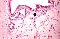
In this slide, the atrophy of the tubules is extending to include the Rete testes (arrow) as well.
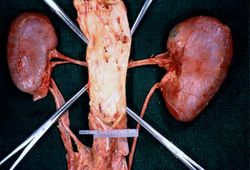
In this gross photograph of kidneys and the abdominal aorta, there is narrowing of the left renal artery at its ostium from the aorta. This atherosclerotic narrowing of the renal artery causes reduced blood pressure in the kidney whose artery is affected. The result is a characteristic Goldblatt’s hypertension syndrome. Note that the kidney with the narrowed renal artery is atrophied compared to the kidney with the normal vessel on the right. What is the cause of the atrophy in this kidney?
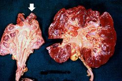
These kidneys were removed from a patient who had blockage of one ureter leading to increased pressure in the renal pelvis. The increased pressure produced hydronephrosis (arrow) in one kidney. What is the cause of atrophy in this case?
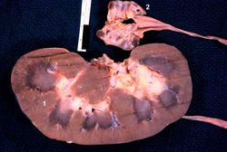
The two kidneys in this slide are from the same patient. One kidney (1) is relatively normal, although increased in size due to compensatory hypertrophy. The other kidney (2) is very small with only rudimentary nodules of renal parenchyma. This kidney had never developed and therefore this process represents hypoplasia. How does one differentiate between atrophy and hypoplasia?
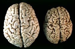
This gross photograph shows a normal brain (left) and a brain from a geriatric patient (right). Note the decreased size, the narrowed gyri, and the widened sulci of the brain from this octogenarian. What is the cause of atrophy in this case?


| |||||
Atherosclerosis is the deposition of lipid into the intima of arteries, resulting in narrowing of the vessel lumen.
Hydronephrosis is dilation of the renal pelvis and atrophy of the cortex due to increase pressure from retained urine.