Difference between revisions of "IPLab:Lab 1:Myocardial Infarction"
Seung Park (talk | contribs) |
(→Myocardial Infarction) |
||
| (17 intermediate revisions by 3 users not shown) | |||
| Line 1: | Line 1: | ||
== Clinical Summary == | == Clinical Summary == | ||
This was a 57-year-old male whose hospital course following abdominal surgery was characterized by progressive deterioration and hypotension. Four days post-operatively, the patient sustained an anterior myocardial infarction and died the next day. | This was a 57-year-old male whose hospital course following abdominal surgery was characterized by progressive deterioration and hypotension. Four days post-operatively, the patient sustained an anterior myocardial infarction and died the next day. | ||
| + | |||
| + | At autopsy the patient's heart weighed 410 grams. Examination of the coronary arteries revealed marked atherosclerotic narrowing of all three vessels with focal occlusion by a thrombus of the left anterior descending artery. Fresh necrosis of the anterior wall of the left ventricle and anterior portion of the septum was present, extending from the endocardium to the inner half of the ventricular wall. | ||
| + | |||
| + | == Images == | ||
| + | <gallery heights="250px" widths="250px"> | ||
| + | File:IPLab1MyocardialInfarction1.jpg|In this gross photograph of the heart from this case, note the area of fresh myocardial infarction (arrows) in the anterior portion of the left ventricle and extending into the anterior portion of the interventricular septum. Note that the walls of the left and right ventricle are slightly thicker than normal. | ||
| + | File:IPLab1MyocardialInfarction2.jpg|This is a low-power photomicrograph of the left ventricular free wall extending from the epicardium (1) to the endocardium (2). The area of infarction is the darker red (hypereosinophilic area) along the subendocardium (3). | ||
| + | File:IPLab1MyocardialInfarction3.jpg|This higher-power photomicrograph shows endocardium on the right side of this image. Directly beneath the endocardium is a pale area consisting of cardiac myocytes exhibiting vacuolar degeneration (1). The area of infarction is visible as a hypereosinophilic area (2) and there is a second zone of vacuolated myocytes (3) between the infarct and the normal myocardium (4). | ||
| + | File:IPLab1MyocardialInfarction4.jpg|This high-power photomicrograph shows the area of infarction on the right (1). There is an area of vacuolated myocytes (2) adjacent to the infarcted myocytes and then normal cardiac muscle to the left (3). | ||
| + | File:IPLab1MyocardialInfarction5.jpg|This high-power photomicrograph shows the endocardium (1) and the area of subendocardial vacuolar degeneration (2). The area of infarction (3) contains some red blood cells. | ||
| + | File:IPLab1MyocardialInfarction6.jpg|This high-power photomicrograph demonstrates the border between the vacuolated subendocardial myocytes (1) and the infarcted myocytes (2). | ||
| + | File:IPLab1MyocardialInfarction7.jpg|This high-power photomicrograph contains normal myocytes (1), vacuolated myocytes (2), and infarcted myocytes (3). | ||
| + | </gallery> | ||
| + | |||
| + | == Virtual Microscopy == | ||
| + | === Myocardial Infarction === | ||
| + | <peir-vm>IPLab1MyocardialInfarction</peir-vm> | ||
| + | |||
| + | === Normal Heart === | ||
| + | <peir-vm>IPLab2Hypertrophy_normal_Heart</peir-vm> | ||
| + | |||
| + | == Study Questions == | ||
| + | * <spoiler text="What type of necrosis is present in this myocardial tissue?">Coagulative necrosis.</spoiler> | ||
| + | * <spoiler text="What are the morphologic characteristics of coagulative necrosis?">Hypereosinophilia, coagulation of cellular proteins, and loss of nuclei (pyknosis, karyolysis, and karyorrhexis).</spoiler> | ||
| + | * <spoiler text="What causes the vacuolar change seen in the tissue adjacent to this infarct and is this change reversible or irreversible injury?">The vacuolar change (hydropic change) seen in myocytes at the edge of an infarct is a REVERSIBLE CHANGE caused by CELLULAR EDEMA. | ||
| + | |||
| + | At the edge of an infarct the oxygen tension is low (hypoxia) so there is a decrease in oxidative metabolism and an increase in anaerobic glycolysis. Since anaerobic glycolysis is less efficient than aerobic metabolism there are lower ATP levels which result in impaired osmotic regulation. In addition, metabolic metabolites accumulate which further increases the intracellular osmotic load and leads to cellular edema.</spoiler> | ||
| + | |||
| + | == Additional Resources == | ||
| + | |||
| + | === Reference === | ||
| + | * [http://emedicine.medscape.com/article/155919-overview eMedicine Medical Library: Myocardial Infarction] | ||
| + | * [http://www.merckmanuals.com/professional/cardiovascular_disorders/coronary_artery_disease/acute_coronary_syndromes_acs.html Merck Manual: Acute Coronary Syndromes] | ||
| + | |||
| + | === Journal Articles === | ||
| + | * Agnew NM, Pennefather SH, Russell GN. [http://onlinelibrary.wiley.com/doi/10.1046/j.1365-2044.2002.02469.x/full Isofluorane and coronary heart disease]. ''Anaesthesia'' 2002; 57(4): 338-347 | ||
| + | |||
| + | === Images === | ||
| + | * [{{SERVER}}/library/index.php?/tags/43-myocardial_infarct PEIR Digital Library: Myocardial Infarct Images] | ||
| + | * [http://library.med.utah.edu/WebPath/CVHTML/CVIDX.html WebPath: Cardiovascular Pathology Images] | ||
| + | |||
| + | == Related IPLab Cases == | ||
| + | * [[IPLab:Lab 3:Acute Myocardial Infarction|Lab 3: Heart: Acute Myocardial Infarction]] | ||
| + | * [[IPLab:Lab 3:Healed Myocardial Infarction|Lab 3: Heart: Healed Myocardial Infarction]] | ||
| + | * [[IPLab:Lab 4:Mural Thrombus|Lab 4: Heart: Mural Thrombus]] | ||
| + | * [[IPLab:Lab 4:Thrombosis|Lab 4: Coronary Artery: Thrombosis]] | ||
| + | * [[IPLab:Lab 4:Pulmonary Congestion and Edema|Lab 4: Lung: Pulmonary Congestion and Edema]] | ||
| + | |||
| + | {{Template:IPLab 1}} | ||
| + | |||
| + | [[Category:IPLab:Lab 1]] | ||
Latest revision as of 00:26, 1 August 2019
Contents
Clinical Summary[edit]
This was a 57-year-old male whose hospital course following abdominal surgery was characterized by progressive deterioration and hypotension. Four days post-operatively, the patient sustained an anterior myocardial infarction and died the next day.
At autopsy the patient's heart weighed 410 grams. Examination of the coronary arteries revealed marked atherosclerotic narrowing of all three vessels with focal occlusion by a thrombus of the left anterior descending artery. Fresh necrosis of the anterior wall of the left ventricle and anterior portion of the septum was present, extending from the endocardium to the inner half of the ventricular wall.
Images[edit]
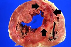
In this gross photograph of the heart from this case, note the area of fresh myocardial infarction (arrows) in the anterior portion of the left ventricle and extending into the anterior portion of the interventricular septum. Note that the walls of the left and right ventricle are slightly thicker than normal.
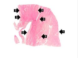
This is a low-power photomicrograph of the left ventricular free wall extending from the epicardium (1) to the endocardium (2). The area of infarction is the darker red (hypereosinophilic area) along the subendocardium (3).
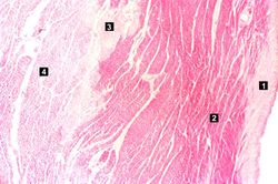
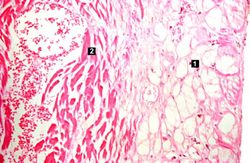
This higher-power photomicrograph shows endocardium on the right side of this image. Directly beneath the endocardium is a pale area consisting of cardiac myocytes exhibiting vacuolar degeneration (1). The area of infarction is visible as a hypereosinophilic area (2) and there is a second zone of vacuolated myocytes (3) between the infarct and the normal myocardium (4).
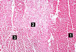
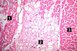
This high-power photomicrograph shows the area of infarction on the right (1). There is an area of vacuolated myocytes (2) adjacent to the infarcted myocytes and then normal cardiac muscle to the left (3).

This high-power photomicrograph shows the endocardium (1) and the area of subendocardial vacuolar degeneration (2). The area of infarction (3) contains some red blood cells.
This high-power photomicrograph demonstrates the border between the vacuolated subendocardial myocytes (1) and the infarcted myocytes (2).
This high-power photomicrograph contains normal myocytes (1), vacuolated myocytes (2), and infarcted myocytes (3).


Virtual Microscopy[edit]
Myocardial Infarction[edit]
Normal Heart[edit]
Study Questions[edit]
Additional Resources[edit]
Reference[edit]
Journal Articles[edit]
- Agnew NM, Pennefather SH, Russell GN. Isofluorane and coronary heart disease. Anaesthesia 2002; 57(4): 338-347
Images[edit]
Related IPLab Cases[edit]
- Lab 3: Heart: Acute Myocardial Infarction
- Lab 3: Heart: Healed Myocardial Infarction
- Lab 4: Heart: Mural Thrombus
- Lab 4: Coronary Artery: Thrombosis
- Lab 4: Lung: Pulmonary Congestion and Edema
| |||||
Myocardial infarction is necrosis of myocardial tissue which occurs as a result of a deprivation of blood supply, and thus oxygen, to the heart tissue. Blockage of blood supply to the myocardium is caused by occlusion of a coronary artery.
Atherosclerosis is the deposition of lipid into the intima of arteries, resulting in narrowing of the vessel lumen.
An occlusion is a blockage.
A thrombus is a solid mass resulting from the aggregation of blood constituents within the vascular system.