From Pathology Education Instructional Resource
Jump to navigation
Jump to search
Images
-
This is a gross photograph of the kidneys from this case. Note that both kidneys contain multiple large cysts (arrows).
-
This is a gross photograph of one of the kidneys from this case next to a normal kidney. This photograph demonstrates how big these polycystic kidneys are compared to a normal kidney.
-
This is a gross photograph of a cut section from one of these polycystic kidneys. Note that the renal parenchyma is almost completely replaced by cystic structures.
-
This is a low-power photomicrograph of an H&E-stained section from this polycystic kidney. Note the large cystic structures (1), the few residual glomeruli (2), and the fibrous connective tissue throughout this section.
-
This is another low-power photomicrograph of an H&E-stained section from this polycystic kidney. Again note the large cystic structures (arrows)and the fibrous connective tissue throughout this section.
-
This is a higher-power photomicrograph of polycystic kidney showing the edge of a large cyst (1). In this section there are numerous tubules and dilated collecting ducts (2) that are filled with the same red proteinaceous material as the larger cysts.
-
This high-power photomicrograph shows abnormal glomeruli (arrows) and some tubules.
-
This is a gross photograph of the liver from this patient. Multiple cysts can be seen on the surface of this liver (arrows).
-
This photomicrograph of liver demonstrates the histologic appearance of these cysts.
-
This is another photomicrograph of liver demonstrating the histologic appearance of these cysts. These cystic structures are associated with the biliary tree.
-
This is a higher-power photomicrograph of liver cyst. These cystic structures are lined by biliary epithelium.
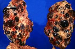 This is a gross photograph of the kidneys from this case. Note that both kidneys contain multiple large cysts (arrows).
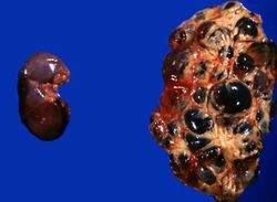 This is a gross photograph of one of the kidneys from this case next to a normal kidney. This photograph demonstrates how big these polycystic kidneys are compared to a normal kidney.
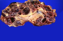 This is a gross photograph of a cut section from one of these polycystic kidneys. Note that the renal parenchyma is almost completely replaced by cystic structures.
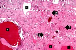 This is a low-power photomicrograph of an H&E-stained section from this polycystic kidney. Note the large cystic structures (1), the few residual glomeruli (2), and the fibrous connective tissue throughout this section.
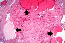 This is another low-power photomicrograph of an H&E-stained section from this polycystic kidney. Again note the large cystic structures (arrows)and the fibrous connective tissue throughout this section.
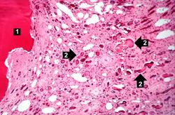 This is a higher-power photomicrograph of polycystic kidney showing the edge of a large cyst (1). In this section there are numerous tubules and dilated collecting ducts (2) that are filled with the same red proteinaceous material as the larger cysts.
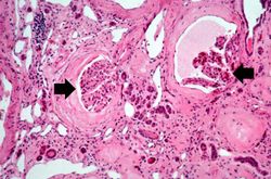 This high-power photomicrograph shows abnormal glomeruli (arrows) and some tubules.
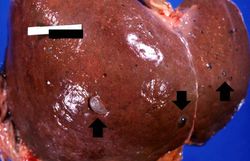 This is a gross photograph of the liver from this patient. Multiple cysts can be seen on the surface of this liver (arrows).
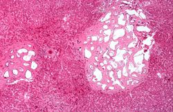 This photomicrograph of liver demonstrates the histologic appearance of these cysts.
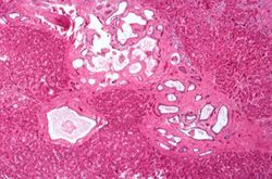 This is another photomicrograph of liver demonstrating the histologic appearance of these cysts. These cystic structures are associated with the biliary tree.
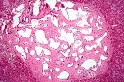 This is a higher-power photomicrograph of liver cyst. These cystic structures are lined by biliary epithelium.


