IPLab:Lab 5:Gout
Jump to navigation
Jump to search
Clinical Summary
This patient was diagnosed with gout approximately 20 years ago. He has had numerous episodes of hot, painful, swollen joints involving the left knee, left ankle, and both first metatarsophalangeal joints. At the time of this admission the patient had increased serum uric acid values. In addition to his painful swollen joints, a gouty tophus was also present on the left arm. After remission of his acute gout flare-up the tophus was surgically removed.
The surgical specimen consisted of an elliptically shaped, mottled, yellow-white irregular hard mass, measuring 8.0 x 5.0 x 2.0 cm. in diameter.
Images
-
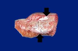 This is a gross photograph of an index finger from a patient with gout. The finger has been sectioned longitudinally to demonstrate the distal interphalangeal joint. Note the white chalky material within and adjacent to the joint (arrows).
 -
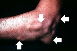 This is a gross photograph of the elbow of this patient. The subcutaneous nodules (arrows) on this arm are tophi caused by gout.
 -
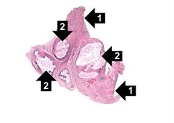 This is a low-power photomicrograph of the tophus removed from the elbow of this patient. Note the fibrous connective tissue (1) and the large foci containing the urate crystals (2) surrounded by the intense chronic inflammatory reaction.
 -
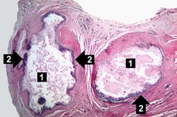 This higher-power photomicrograph of the tophus demonstrates the collections of urate crystals (1) and the inflammatory cells at the edge of these foci (2).
 -
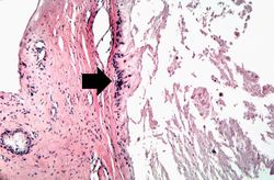 This is a higher-power photomicrograph of the edge of the tophus. Most of the urate crystals dissolve away during processing. The inflammatory cells at the edge of these foci are clearly visible (arrow).
 -
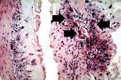 This is a high-power photomicrograph of the edge of the tophus. The character of the intense chronic inflammatory cell reaction is evident and note the presence of giant cells within this inflammatory cell reaction (arrows).
 -
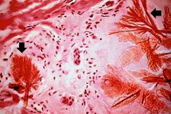 This is a photomicrograph of a tophus that was fixed in alcohol prior to histologic processing. The alcohol fixation preserves the water soluble urate crystals within the tissue. Note the urate crystals visible in this photomicrograph (arrows). Also note the chronic inflammatory reaction in the background.
 -
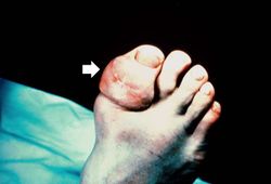


Virtual Microscopy
Study Questions
Additional Resources
Reference
- eMedicine Medical Library: Gout and Pseudogout
- American Academy of Family Physicians: Gout and Hyperuricemia
Journal Articles
- Harris MD, Siegel LB, Alloway JA. Gout and hyperuricemia. Am Fam Physician 1999 Feb 15;59(4):925-34.
- Pittman JR, Bross MH. Diagnosis and management of gout. Am Fam Physician 1999 Apr 1;59(7):1799-806, 1810.
- Qaseem A, Harris RP, Forciea MA. Management of Acute and Recurrent Gout. Ann Intern Med 2016 Nov 1 10;7326/M16-0570.
Images
Related IPLab Cases
| |||||