From Pathology Education Instructional Resource
Jump to navigation
Jump to search
Images
-
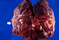
This is a gross photograph of lung demonstrating areas of fibrosis on the pleural surface (arrow).
-
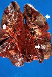
This is a gross photograph of cut sections of lung. There are several areas of fibrosis (arrows) within the lung parenchyma.
-
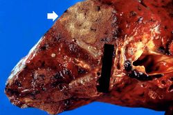
This is a gross photograph showing a closer view of a cut section of lung. An area of fibrosis (arrow) is evident in this photograph.
-
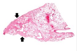
This is a low-power photomicrograph of lung section. Note the thickening of the alveolar septa (arrows).
-
This is a higher-power photomicrograph of lung section. Note the thickening of the alveolar septa (1) and accumulations of
anthracotic pigment (2).
-
This is another high-power photomicrograph of lung section showing the thickening of the alveolar septa (arrows) and accumulations of black
anthracotic pigment.
-
This high-power photomicrograph of lung section shows the thickening of the alveolar septum (arrows) by fibrous connective tissue.
-
This is a photomicrograph of a trichrome-stained section of lung demonstrating the extensive fibrosis throughout this section (green-blue stained material is fibrous connective tissue).
-
This is a photomicrograph of an area of tissue exhibiting diffuse fibrosis and thickening of the alveolar septa.
-
This is another high-power photomicrograph of an area of tissue with diffuse fibrosis and thickening of the alveolar septa. There are also accumulations of
anthracotic pigment in this area (arrows).
-
This medium-power photomicrograph shows fibrosis and severe intimal changes in blood vessels (arrows).
-
This high-power photomicrograph shows intimal changes (arrows) in this blood vessel in the lung.
-
This is a high-power photomicrograph of a
recanalized blood vessel in the lung. Notice the
anthracotic pigment adjacent to the vessel (arrows).
 This is a gross photograph of lung demonstrating areas of fibrosis on the pleural surface (arrow).
 This is a gross photograph of cut sections of lung. There are several areas of fibrosis (arrows) within the lung parenchyma.
 This is a gross photograph showing a closer view of a cut section of lung. An area of fibrosis (arrow) is evident in this photograph.
 This is a low-power photomicrograph of lung section. Note the thickening of the alveolar septa (arrows).
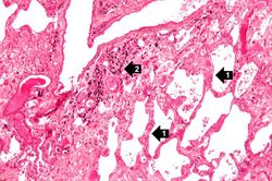 This is a higher-power photomicrograph of lung section. Note the thickening of the alveolar septa (1) and accumulations of anthracotic pigment (2).
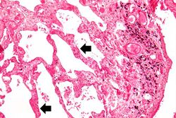 This is another high-power photomicrograph of lung section showing the thickening of the alveolar septa (arrows) and accumulations of black anthracotic pigment.
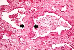 This high-power photomicrograph of lung section shows the thickening of the alveolar septum (arrows) by fibrous connective tissue.
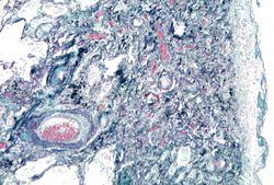 This is a photomicrograph of a trichrome-stained section of lung demonstrating the extensive fibrosis throughout this section (green-blue stained material is fibrous connective tissue).
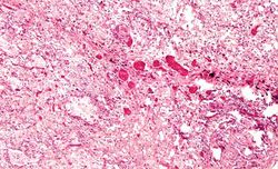 This is a photomicrograph of an area of tissue exhibiting diffuse fibrosis and thickening of the alveolar septa.
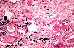 This is another high-power photomicrograph of an area of tissue with diffuse fibrosis and thickening of the alveolar septa. There are also accumulations of anthracotic pigment in this area (arrows).
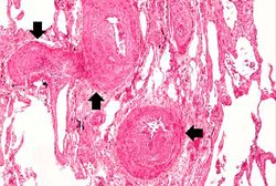 This medium-power photomicrograph shows fibrosis and severe intimal changes in blood vessels (arrows).
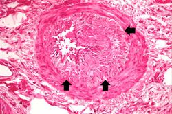 This high-power photomicrograph shows intimal changes (arrows) in this blood vessel in the lung.
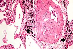 This is a high-power photomicrograph of a recanalized blood vessel in the lung. Notice the anthracotic pigment adjacent to the vessel (arrows).


