From Pathology Education Instructional Resource
Jump to navigation
Jump to search
Images
-
This gross photograph of the liver from this patient demonstrates the pale areas of necrosis (arrows).
-
In this photomicrograph of the liver from this patient there are areas of hemorrhagic necrosis (arrows).
-
This is another example of the areas of hemorrhagic necrosis (arrows).
-
This is a medium-power photomicrograph of the areas of hemorrhagic necrosis. Note the coagulation necrosis and hemorrhage in this area. Viable hepatocytes can be seen along the edge of this lesion.
-
This is a high-power photomicrograph of the edge of the necrosis. Again, note the coagulation necrosis and hemorrhage in this area. The viable hepatocytes at the edge of this necrosis are vacuolated (arrows).
-
Photograph taken at autopsy demonstrating the severe necrosis of the skin of foot.
-
This low-power photomicrograph of the skin from this patient shows a blister and numerous thrombosed vessels (arrows) in the dermis.
-
This is a higher-power photomicrograph of the skin from this patient showing a blister and the thrombosed small vessels (arrows) in the dermis.
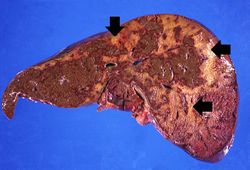 This gross photograph of the liver from this patient demonstrates the pale areas of necrosis (arrows).
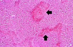 In this photomicrograph of the liver from this patient there are areas of hemorrhagic necrosis (arrows).
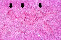 This is another example of the areas of hemorrhagic necrosis (arrows).
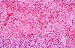 This is a medium-power photomicrograph of the areas of hemorrhagic necrosis. Note the coagulation necrosis and hemorrhage in this area. Viable hepatocytes can be seen along the edge of this lesion.
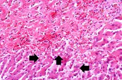 This is a high-power photomicrograph of the edge of the necrosis. Again, note the coagulation necrosis and hemorrhage in this area. The viable hepatocytes at the edge of this necrosis are vacuolated (arrows).
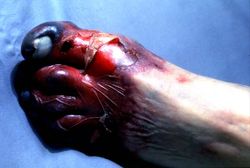 Photograph taken at autopsy demonstrating the severe necrosis of the skin of foot.
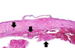 This low-power photomicrograph of the skin from this patient shows a blister and numerous thrombosed vessels (arrows) in the dermis.
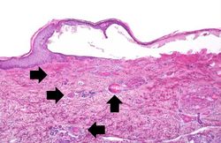 This is a higher-power photomicrograph of the skin from this patient showing a blister and the thrombosed small vessels (arrows) in the dermis.


