IPLab:Lab 5:α1 Antitrypsin Deficiency: Difference between revisions
Jump to navigation
Jump to search
No edit summary |
No edit summary |
||
| Line 5: | Line 5: | ||
File:IPLab5Antitrypsin3.jpg|This is a gross photograph of the bronchi and lungs. Note the hemorrhage in the bronchi and in the lung parenchyma. | File:IPLab5Antitrypsin3.jpg|This is a gross photograph of the bronchi and lungs. Note the hemorrhage in the bronchi and in the lung parenchyma. | ||
File:IPLab5Antitrypsin4.jpg|This is a low-power photomicrograph from an area of the lung without significant hemorrhage. The enlarged, emphysematous air spaces are easily appreciated. | File:IPLab5Antitrypsin4.jpg|This is a low-power photomicrograph from an area of the lung without significant hemorrhage. The enlarged, emphysematous air spaces are easily appreciated. | ||
File:IPLab5Antitrypsin5.jpg|This is a | File:IPLab5Antitrypsin5.jpg|This is a low-power photomicrograph from an area of the lung without significant hemorrhage. The enlarged, emphysematous air spaces are easily appreciated. | ||
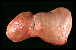
File:IPLab5Antitrypsin6.jpg| | File:IPLab5Antitrypsin6.jpg| | ||
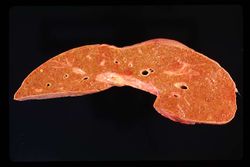
File:IPLab5Antitrypsin7.jpg| | File:IPLab5Antitrypsin7.jpg| | ||
Revision as of 13:19, 19 August 2013
Images
-
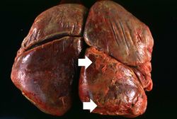 This is a gross photograph of the lungs from this case. The rough friable material on the surface of the lung (arrows) is fibrinous exudate and fibrous tissue. This reaction on the surface of the lung is due to the recent surgery. The emphysematous changes are not easily appreciated in this photograph.
 -
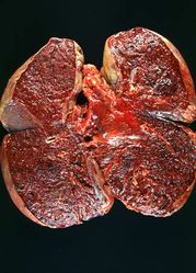 This is a gross photograph of the cut sections of lung from this case. The lung parenchyma is markedly hemorrhagic and consolidated. Again the hemorrhage makes it difficult to appreciate the emphysematous changes.
 -
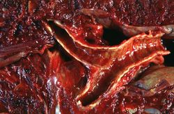 This is a gross photograph of the bronchi and lungs. Note the hemorrhage in the bronchi and in the lung parenchyma.
 -
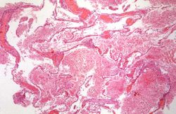 This is a low-power photomicrograph from an area of the lung without significant hemorrhage. The enlarged, emphysematous air spaces are easily appreciated.
 -
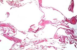 This is a low-power photomicrograph from an area of the lung without significant hemorrhage. The enlarged, emphysematous air spaces are easily appreciated.
 -
-
-
-
-
-
-


| |||||