IPLab:Lab 2:Metaplasia: Difference between revisions
No edit summary |
Seung Park (talk | contribs) |
||
| Line 35: | Line 35: | ||
=== Images === | === Images === | ||
* [ | * [{{SERVER}}/library/index.php?/tags/148-bladder/11-urinary PEIR Digital Library: Bladder Images] | ||
* [http://library.med.utah.edu/WebPath/RENAHTML/RENALIDX.html WebPath: Renal Pathology Images] | * [http://library.med.utah.edu/WebPath/RENAHTML/RENALIDX.html WebPath: Renal Pathology Images] | ||
Revision as of 01:24, 30 August 2013
Clinical Summary
This 30-year-old black male was born with a meningocele which was repaired in childhood. Despite repair of the meningocele, this patient continued to have a neurogenic bladder.
Six months previous to this admission, the patient passed a single renal calculus but intravenous pyelogram (IVP) showed left nephrolithiasis (a 1 cm stone in the lower lobe). For 6 months the patient had to be catheterized each day for 6 hours. However, he continued to complain of hesitancy and urgency and suffered occasional urinary tract infections.
On this admission, a culture of the patient's urine specimen grew Gram-negative bacilli (Proteus species). Cystoscopy showed heavy trabeculation of the bladder with early diverticula and the left ureteral orifice showed squamous metaplasia. The lower pole of the left kidney was removed surgically, following which the patient recovered and was discharged on antibiotics.
Images
-
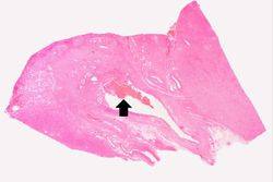 This is a low-power photomicrograph showing the full cortical and medullary thickness of the kidney. Note that there is a dilated calyx containing some red blood cells in the center of the section (arrow). The cortex is markedly thin and has severe lesions of degeneration and atrophy, although these are hard to appreciate at this low magnification.
 -
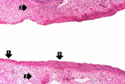 This high-power photomicrograph demonstrates the transitional epithelium lining the renal calyx (1) and the junction (transition zone) to a thicker hyperplastic epithelium (2). Note the inflammatory cells and increased vascular response in the stromal tissue (3) lying beneath the normal transitional epithelium.
 -
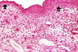 A higher-power view shows the junction of normal epithelium (1) with hyperplastic transitional epithelium (2). Note the inflammatory cells in the subepithelial tissue.
 -
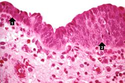 This is a higher-power photomicrograph of the junction of normal epithelium (1) with hyperplastic transitional epithelium (2).
 -
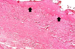 In areas adjacent to the normal transitional epithelium, there are areas of epithelium (arrows) where the epithelial cells have the character of normal squamous epithelium as found in the dermis. However, squamous epithelium is not normal in the renal pelvis. This adaptive change is referred to as squamous metaplasia.
 -
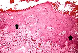 A high-power photomicrograph of the squamous epithelium shows inflammatory cells in the subepithelial tissue and the formation of keratinized epithelium (arrows).
 -
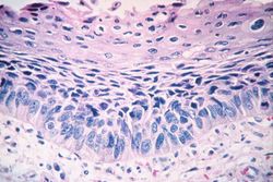 This is a photomicrograph of the trachea from a smoker. Note that the columnar ciliated epithelium has been replaced by squamous epithelium.


Study Questions
- <spoiler text="What is a neurogenic bladder and what complications may result from this condition?">A neurogenic bladder is any condition or dysfunction of the urinary bladder caused by a lesion of the central or peripheral nervous system. Complications primarily include stasis, infection, and stone formation.</spoiler>
- <spoiler text="What factors in this case predisposed to nephrolithiasis?">Stasis and infection.</spoiler>
- <spoiler text="Does this metaplastic process predispose to neoplasia?">Yes.</spoiler>
Additional Resources
Reference
- eMedicine Medical Library: Nephrolithiasis
- eMedicine Medical Library: Neurogenic Bladder
- Merck Manual: Urinary Calculi
- Merck Manual: Neurogenic Bladder
- Merck Manual: Approach to the Genitourinary Patient
Journal Articles
- Pak CY, Sakhaee K, Peterson RD, Poindexter JR, Frawley WH. Biochemical profile of idiopathic uric acid nephrolithiasis. Kidney Int 2001 Aug;60(2):757-61.
Images
| |||||