IPLab:Lab 5:Gaucher Disease: Difference between revisions
Seung Park (talk | contribs) No edit summary |
Seung Park (talk | contribs) No edit summary |
||
| Line 25: | Line 25: | ||
The third pattern, type III, is usually somewhat intermediate between types I and II. These patients are usually juveniles and have systemic involvement like in type I but they have progressive central nervous system disease that usually begins in the second or third decade of life.</spoiler> | The third pattern, type III, is usually somewhat intermediate between types I and II. These patients are usually juveniles and have systemic involvement like in type I but they have progressive central nervous system disease that usually begins in the second or third decade of life.</spoiler> | ||
* <spoiler text="What type of Gaucher disease did this patient likely have?">This was an adult who had pancytopenia and aseptic necrosis of bone. This is consistent with type I. In type I symptoms and signs first appear in adult life and are related to splenomegaly or to bone involvement by Gaucher cells. These patients have pancytopenia or thrombocytopenia secondary to hypersplenism and pathologic fractures and bone pain due to expansion of the marrow space by Gaucher cells.</spoiler> | * <spoiler text="What type of Gaucher disease did this patient likely have?">This was an adult who had pancytopenia and aseptic necrosis of bone. This is consistent with type I. In type I symptoms and signs first appear in adult life and are related to splenomegaly or to bone involvement by Gaucher cells. These patients have pancytopenia or thrombocytopenia secondary to hypersplenism and pathologic fractures and bone pain due to expansion of the marrow space by Gaucher cells.</spoiler> | ||
== Additional Resources == | |||
=== Reference === | |||
* [http://emedicine.medscape.com/article/944157-overview eMedicine Medical Library: Gaucher Disease] | |||
* [http://www.merckmanuals.com/professional/hematology_and_oncology/spleen_disorders/splenomegaly.html Merck Manual: Splenomegaly] | |||
* [www.merckmanuals.com/professional/pediatrics/inherited_disorders_of_metabolism/lysosomal_storage_disorders.html Merck Manual: Lysosomal Storage Disorders] | |||
=== Journal Articles === | |||
* Barone R, Pavone V, Nigro F, Chabàs A, Fiumara A. [http://www.ncbi.nlm.nih.gov/pubmed/10792292 Extraordinary bone involvement in a gaucher disease type I patient]. ''Br J Haematol'' 2000 Mar;108(4):838-41. | |||
=== Images === | |||
* [http://peir.path.uab.edu/library/index.php?/tags/1670-gaucher_cell PEIR Digital Library: Gaucher Cell Images] | |||
* [http://library.med.utah.edu/WebPath/HEMEHTML/HEMEIDX.html Webpath: Hematopathology] | |||
{{IPLab 5}} | {{IPLab 5}} | ||
[[Category: IPLab:Lab 5]] | [[Category: IPLab:Lab 5]] | ||
Revision as of 15:46, 22 August 2013
Clinical Summary =
This 23-year-old black female delivered a stillborn infant by Caesarean section, following which she experienced excessive uterine bleeding including the passage of blood clots. Further study revealed an enlarged spleen and thrombocytopenia (platelet count 58,000). A splenectomy was performed with an uneventful postoperative course other than persistent pain in her right leg (the right hip had been fractured 2 years earlier). Subsequently she developed aseptic necrosis of the left femoral head requiring a prosthetic replacement. Microscopic evaluation of the bone removed at the time of surgery revealed Gaucher cells in the marrow space.
Autopsy Findings
The surgical specimen was a 935-gram spleen. Its surface was pale with an area of bluish discoloration. The cut surface revealed the same pale appearance.
Images
-
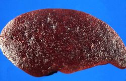 This is a gross photograph of spleen from this case. The spleen is enlarged and the surface is finely granular.
 -
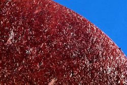 This is a cut section of spleen from this case. Again note the fine granular appearance to the tissue.
 -
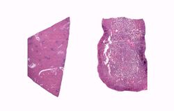 This is a low-power photomicrograph of normal spleen (left) and the spleen from this case (right). The loose appearance of the tissue in the Gaucher spleen is due to artifactual loss of tissue during histologic processing.
 -
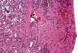 This is a photomicrograph of the spleen from this case. There is very little if any white pulp evident in this section.
 -
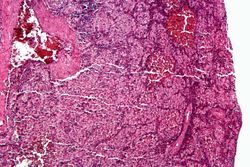 This is a higher-power photomicrograph of the spleen from this case. Again there is no white pulp and the red pulp is filled with large eosinophilic cells.
 -
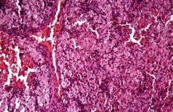 This is another high-power photomicrograph of the spleen from this case. At this power it is easier to see the large eosinophilic cells.
 -
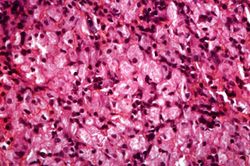 This is another high-power photomicrograph of the spleen from this case. At this high power individual cells can be better appreciated.
 -
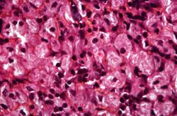 This is a higher-power photomicrograph of the spleen from this case. At this higher power individual cells can be better appreciated and the fibrillar nature of the eosinophilic cytoplasmic material can be seen.


Study Questions
Additional Resources
Reference
- eMedicine Medical Library: Gaucher Disease
- Merck Manual: Splenomegaly
- [www.merckmanuals.com/professional/pediatrics/inherited_disorders_of_metabolism/lysosomal_storage_disorders.html Merck Manual: Lysosomal Storage Disorders]
Journal Articles
- Barone R, Pavone V, Nigro F, Chabàs A, Fiumara A. Extraordinary bone involvement in a gaucher disease type I patient. Br J Haematol 2000 Mar;108(4):838-41.
Images
| |||||