IPLab:Lab 5:Neurofibromatosis: Difference between revisions
Jump to navigation
Jump to search
Created page with "== Images == <gallery heights="250px" widths="250px"> File:IPLab5Neurofibromatosis1.jpg|This photograph, taken at autopsy, demonstrates the distribution of neurofibromas on th..." |
No edit summary |
||
| Line 5: | Line 5: | ||
File:IPLab5Neurofibromatosis3.jpg|This is a closer view of neurofibromas on the skin. | File:IPLab5Neurofibromatosis3.jpg|This is a closer view of neurofibromas on the skin. | ||
File:IPLab5Neurofibromatosis4.jpg|This is a low-power photomicrograph of a subcutaneous neurofibroma (1). Note the increased pigmentation in the skin (2). | File:IPLab5Neurofibromatosis4.jpg|This is a low-power photomicrograph of a subcutaneous neurofibroma (1). Note the increased pigmentation in the skin (2). | ||
File:IPLab5Neurofibromatosis5.jpg|This is a higher-power photomicrograph of the neurofibroma (1) with the overlying skin (2). | |||
File:IPLab5Neurofibromatosis6.jpg|This is a higher-power photomicrograph of the neurofibroma demonstrating the loose pattern of elongated cells making up the tumor mass. | File:IPLab5Neurofibromatosis6.jpg|This is a higher-power photomicrograph of the neurofibroma demonstrating the loose pattern of elongated cells making up the tumor mass. | ||
File:IPLab5Neurofibromatosis7.jpg|This higher-power photomicrograph of the neurofibroma shows more clearly the elongated cells (primarily Schwann cells) that make up this tumor. | File:IPLab5Neurofibromatosis7.jpg|This higher-power photomicrograph of the neurofibroma shows more clearly the elongated cells (primarily Schwann cells) that make up this tumor. | ||
Revision as of 12:23, 19 August 2013
Images
-
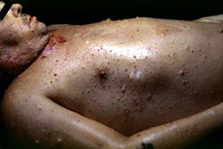 This photograph, taken at autopsy, demonstrates the distribution of neurofibromas on the skin of this patient.
 -
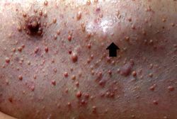 This is another view taken at autopsy demonstrating the neurofibromas. Some lesions can be seen as subcutaneous swellings (arrow) and others form pedunculated masses. Most are hyperpigmented.
 -
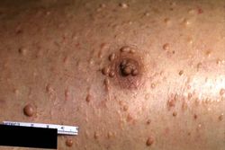 This is a closer view of neurofibromas on the skin.
 -
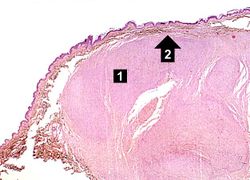 This is a low-power photomicrograph of a subcutaneous neurofibroma (1). Note the increased pigmentation in the skin (2).
 -
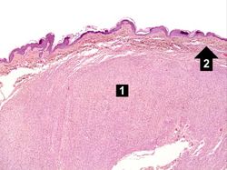 This is a higher-power photomicrograph of the neurofibroma (1) with the overlying skin (2).
 -
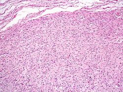 This is a higher-power photomicrograph of the neurofibroma demonstrating the loose pattern of elongated cells making up the tumor mass.
 -
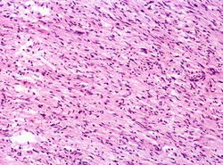 This higher-power photomicrograph of the neurofibroma shows more clearly the elongated cells (primarily Schwann cells) that make up this tumor.
 -
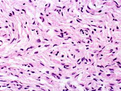 This is a high-power photomicrograph of the cells in the neurofibroma.


| |||||