IPLab:Lab 2:Metaplasia: Difference between revisions
Jump to navigation
Jump to search
Created page with "== Images == <gallery heights="250px" widths="250px"> File:IPLab2Metaplasia1.jpg|This is a low-power photomicrograph showing the full cortical and medullary thickness of the k..." |
No edit summary |
||
| Line 2: | Line 2: | ||
<gallery heights="250px" widths="250px"> | <gallery heights="250px" widths="250px"> | ||
File:IPLab2Metaplasia1.jpg|This is a low-power photomicrograph showing the full cortical and medullary thickness of the kidney. Note that there is a dilated calyx containing some red blood cells in the center of the section (arrow). The cortex is markedly thin and has severe lesions of degeneration and atrophy, although these are hard to appreciate at this low magnification. | File:IPLab2Metaplasia1.jpg|This is a low-power photomicrograph showing the full cortical and medullary thickness of the kidney. Note that there is a dilated calyx containing some red blood cells in the center of the section (arrow). The cortex is markedly thin and has severe lesions of degeneration and atrophy, although these are hard to appreciate at this low magnification. | ||
File:IPLab2Metaplasia2.jpg| | File:IPLab2Metaplasia2.jpg|This high-power photomicrograph demonstrates the transitional epithelium lining the renal calyx (1) and the junction (transition zone) to a thicker hyperplastic epithelium (2). Note the inflammatory cells and increased vascular response in the stromal tissue (3) lying beneath the normal transitional epithelium. | ||
File:IPLab2Metaplasia3.jpg|A higher-power view shows the junction of normal epithelium (1) with hyperplastic transitional epithelium (2). Note the inflammatory cells in the subepithelial tissue. | File:IPLab2Metaplasia3.jpg|A higher-power view shows the junction of normal epithelium (1) with hyperplastic transitional epithelium (2). Note the inflammatory cells in the subepithelial tissue. | ||
File:IPLab2Metaplasia4.jpg|This is a higher-power photomicrograph of the junction of normal epithelium (1) with hyperplastic transitional epithelium (2). | File:IPLab2Metaplasia4.jpg|This is a higher-power photomicrograph of the junction of normal epithelium (1) with hyperplastic transitional epithelium (2). | ||
Revision as of 10:47, 19 August 2013
Images
-
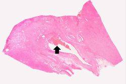 This is a low-power photomicrograph showing the full cortical and medullary thickness of the kidney. Note that there is a dilated calyx containing some red blood cells in the center of the section (arrow). The cortex is markedly thin and has severe lesions of degeneration and atrophy, although these are hard to appreciate at this low magnification.
 -
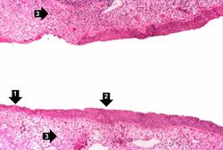 This high-power photomicrograph demonstrates the transitional epithelium lining the renal calyx (1) and the junction (transition zone) to a thicker hyperplastic epithelium (2). Note the inflammatory cells and increased vascular response in the stromal tissue (3) lying beneath the normal transitional epithelium.
 -
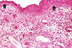 A higher-power view shows the junction of normal epithelium (1) with hyperplastic transitional epithelium (2). Note the inflammatory cells in the subepithelial tissue.
 -
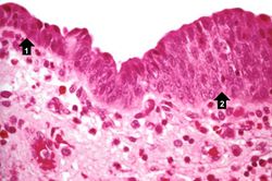 This is a higher-power photomicrograph of the junction of normal epithelium (1) with hyperplastic transitional epithelium (2).
 -
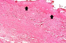 In areas adjacent to the normal transitional epithelium, there are areas of epithelium (arrows) where the epithelial cells have the character of normal squamous epithelium as found in the dermis. However, squamous epithelium is not normal in the renal pelvis. This adaptive change is referred to as squamous metaplasia.
 -
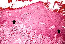 A high-power photomicrograph of the squamous epithelium shows inflammatory cells in the subepithelial tissue and the formation of keratinized epithelium (arrows).
 -
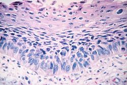 This is a photomicrograph of the trachea from a smoker. Note that the columnar ciliated epithelium has been replaced by squamous epithelium.


| |||||