IPLab:Lab 2:Hypertrophy: Difference between revisions
Seung Park (talk | contribs) No edit summary |
Seung Park (talk | contribs) No edit summary |
||
| Line 16: | Line 16: | ||
File:IPLab2Hypertrophy6.jpg|This gross photograph shows an example of normal physiologic hypertrophy. The organs shown are an open uterus (1), cervix (2) and vagina (3), both ovaries (4) and both kidneys (5) from a woman who died shortly after normal delivery from causes unrelated to childbirth. Note the marked thickening of the uterine wall due to smooth muscle cell hypertrophy. | File:IPLab2Hypertrophy6.jpg|This gross photograph shows an example of normal physiologic hypertrophy. The organs shown are an open uterus (1), cervix (2) and vagina (3), both ovaries (4) and both kidneys (5) from a woman who died shortly after normal delivery from causes unrelated to childbirth. Note the marked thickening of the uterine wall due to smooth muscle cell hypertrophy. | ||
</gallery> | </gallery> | ||
== Study Questions == | |||
* <spoiler text="Is this case of myocardial hypertrophy an example of physiologic or pathologic hypertrophy?">Pathologic.</spoiler> | |||
* <spoiler text="What are some factors that lead to the decompensation of this heart?">The exact mechanism for why hypertrophied hearts go on to develop congestive heart failure is not known. However, some contributing factors may include: | |||
# limitation of the vascular supply to the enlarged myocardial fibers, | |||
# diminished oxidative capabilities of the mitochondria, | |||
# alterations in protein synthesis & degradation, or | |||
# alterations in the cytoskeleton.</spoiler> | |||
== Additional Resources == | |||
=== Reference === | |||
* [http://emedicine.medscape.com/article/890068-overview eMedicine Medical Library: Pediatric Hypertrophic Cardiomyopathy] | |||
* [http://emedicine.medscape.com/article/163062-overview eMedicine Medical Library: Heart Failure] | |||
* [http://www.merckmanuals.com/professional/cardiovascular_disorders/cardiomyopathies/hypertrophic_cardiomyopathy.html Merck Manual: Hypertrophic Cardiomyopathy] | |||
* [http://www.merckmanuals.com/professional/cardiovascular_disorders/heart_failure/heart_failure_hf.html Merck Manual: Heart Failure] | |||
{{Template:IPLab 2}} | {{Template:IPLab 2}} | ||
Revision as of 20:28, 18 August 2013
Clinical Summary
This 68-year-old man initially sought medical advice five years prior to his death. His symptoms at that time were exercise intolerance and occasional peripheral edema. He gave a history of a "heart murmur" that was diagnosed 25 years ago during an employment physical. No follow up care had been given for this murmur.
The patient's terminal admission was for signs of severe heart failure--the patient had marked peripheral edema and shortness of breath and chest x-ray revealed significant cardiac enlargement and pulmonary edema with bilateral pleural effusions. He sustained a cardiac arrest shortly after admission and could not be resuscitated.
Autopsy Findings
Autopsy disclosed a markedly enlarged heart weighing 650 grams and having dilated chambers. The aortic valve was calcified and showed evidence of stenosis and insufficiency. The coronary arteries were narrowed 60 to 70% by atherosclerosis. No acute coronary occlusions were found and there was no evidence of myocardial infarction.
Images
-
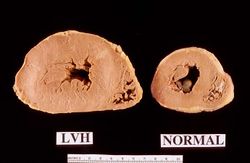 This is a gross photograph of a cross section of a normal human heart taken at autopsy (right) and the heart from this case, which demonstrates concentric hypertrophy of the left ventricular wall. Note the marked thickening of the left ventricular wall. There is also moderate thickening of the right ventricular wall.
 -
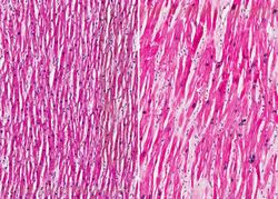 This low-power photomicrograph shows normal myocardium (left) compared to hypertrophied myocardium (right).
 -
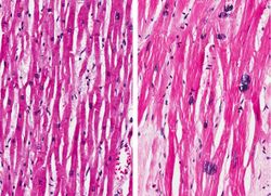 Normal myocardium (left) is compared here to hypertrophied myocardium (right). The muscle fibers are thicker and the nuclei are larger and darker in the hypertrophied myocardium.The clear spaces between the muscle fibers are due to processing artifacts and are not present during life.
 -
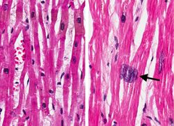 Normal myocardium (left) is compared to hypertrophied myocardium (right). This high power view demonstrates the large dark nuclei (arrow) found in hypertrophied cardiac muscle cells. Polyploidy is a common feature in cardiac hypertrophy. Also note the increased size (thickness) of the individual cardiac muscle cell on the right compared to normal cardiac myocytes (left).
 -
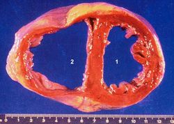 This autopsy specimen was taken from another patient who had cardiac hypertrophy and congestive heart failure that resulted in dilation of the cardiac chambers. This heart was markedly enlarged (700 grams) but the congestive failure leads to dilation of the chambers and thinning of the wall. Note the marked dilation of both the left (1) and right (2) ventricular chambers.
 -
 This gross photograph shows an example of normal physiologic hypertrophy. The organs shown are an open uterus (1), cervix (2) and vagina (3), both ovaries (4) and both kidneys (5) from a woman who died shortly after normal delivery from causes unrelated to childbirth. Note the marked thickening of the uterine wall due to smooth muscle cell hypertrophy.


Study Questions
Additional Resources
Reference
- eMedicine Medical Library: Pediatric Hypertrophic Cardiomyopathy
- eMedicine Medical Library: Heart Failure
- Merck Manual: Hypertrophic Cardiomyopathy
- Merck Manual: Heart Failure
| |||||