IPLab:Lab 12:Radiation Fibrosis: Difference between revisions
No edit summary |
Seung Park (talk | contribs) No edit summary |
||
| Line 35: | Line 35: | ||
Subtle genetic injuries, such as DNA strand breaks, are responsible for translocations and deletions. These changes lead to the mutagenic, teratogenic, and carcinogenic potentials of ionizing radiation that become evident many years after the radiation exposure. During this long time interval, sequential mitotic divisions are occurring that will ultimately lead to these untoward consequences. This phenomenon is known as radiation "latency."</spoiler> | Subtle genetic injuries, such as DNA strand breaks, are responsible for translocations and deletions. These changes lead to the mutagenic, teratogenic, and carcinogenic potentials of ionizing radiation that become evident many years after the radiation exposure. During this long time interval, sequential mitotic divisions are occurring that will ultimately lead to these untoward consequences. This phenomenon is known as radiation "latency."</spoiler> | ||
== Additional Resources == | |||
=== Reference === | |||
=== Journal Articles === | |||
=== Images === | |||
== Related IPLab Cases == | |||
{{IPLab 12}} | {{IPLab 12}} | ||
[[Category: IPLab:Lab 12]] | [[Category: IPLab:Lab 12]] | ||
Revision as of 23:26, 22 August 2013
Clinical Summary
This 60-year-old white female had developed retraction of her left nipple six years earlier, at which time breast carcinoma was found. A radical mastectomy was performed. Examination of the surgical specimens showed metastases in regional lymph nodes and local irradiation was thus administered. Two years later, carcinoma of the right breast was found. Following a modified mastectomy, more irradiation was given. A year later the patient developed recurrences for which chemotherapy (cytoxan and adriamycin) was given. After a two year period without problems, the patient developed decreased exercise tolerance, dyspnea on exertion, shortness of breath, paroxysmal nocturnal dyspnea, and orthopnea increasing in severity over 10 days. Chest examination revealed decreased breath sounds with dullness over the left base. Chest x-ray showed a globose cardiac silhouette and left pleural effusion. A pericardiectomy was done because of suspected cardiac tamponade; however, the patient died soon after the operation.
Autopsy Findings
There was metastatic carcinoma in the pericardium, chest wall, diaphragm, both lungs, and mediastinal lymph nodes. Severe nonobstructive cardiomyopathy, probably secondary to adriamycin, was found. Areas of pleural thickening with adhesions and interstitial fibrosis were found involving the anterior aspect of both lungs.
Images
-
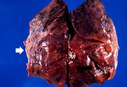 This is a gross photograph of lung demonstrating areas of fibrosis on the pleural surface (arrow).
 -
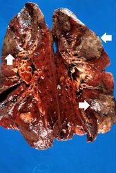 This is a gross photograph of cut sections of lung. There are several areas of fibrosis (arrows) within the lung parenchyma.
 -
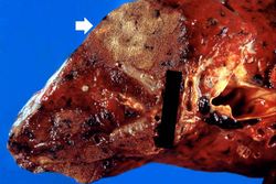 This is a gross photograph showing a closer view of a cut section of lung. An area of fibrosis (arrow) is evident in this photograph.
 -
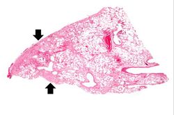 This is a low-power photomicrograph of lung section. Note the thickening of the alveolar septa (arrows).
 -
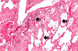 This is a higher-power photomicrograph of lung section. Note the thickening of the alveolar septa (1) and accumulations of anthracotic pigment (2).
 -
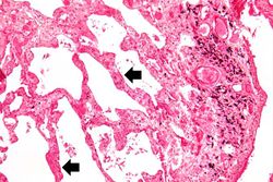 This is another high-power photomicrograph of lung section showing the thickening of the alveolar septa (arrows) and accumulations of black anthracotic pigment.
 -
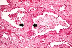 This high-power photomicrograph of lung section shows the thickening of the alveolar septum (arrows) by fibrous connective tissue.
 -
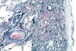 This is a photomicrograph of a trichrome-stained section of lung demonstrating the extensive fibrosis throughout this section (green-blue stained material is fibrous connective tissue).
 -
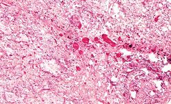 This is a photomicrograph of an area of tissue exhibiting diffuse fibrosis and thickening of the alveolar septa.
 -
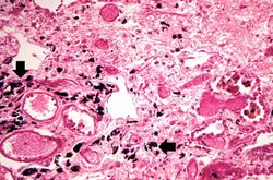 This is another high-power photomicrograph of an area of tissue with diffuse fibrosis and thickening of the alveolar septa. There are also accumulations of anthracotic pigment in this area (arrows).
 -
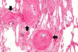 This medium-power photomicrograph shows fibrosis and severe intimal changes in blood vessels (arrows).
 -
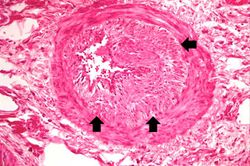 This high-power photomicrograph shows intimal changes (arrows) in this blood vessel in the lung.
 -
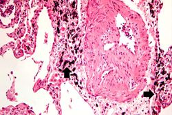 This is a high-power photomicrograph of a recanalized blood vessel in the lung. Notice the anthracotic pigment adjacent to the vessel (arrows).


Study Questions
Additional Resources
Reference
Journal Articles
Images
Related IPLab Cases
| |||||