IPLab:Lab 2:Hyperplasia: Difference between revisions
No edit summary |
No edit summary |
||
| Line 26: | Line 26: | ||
* <spoiler text="What is the age-specific incidence of nodular hyperplasia in males?">The incidence of nodular hyperplasia is: | * <spoiler text="What is the age-specific incidence of nodular hyperplasia in males?">The incidence of nodular hyperplasia is: | ||
20% in men 40 years of age | * 20% in men 40 years of age | ||
30% in men 50 years of age | * 30% in men 50 years of age | ||
70% in men 60 years of age | * 70% in men 60 years of age | ||
90% in men 70 years of age</spoiler> | * 90% in men 70 years of age</spoiler> | ||
* <spoiler text="Does nodular hyperplasia predispose to adenocarcinoma?">No.</spoiler> | * <spoiler text="Does nodular hyperplasia predispose to adenocarcinoma?">No.</spoiler> | ||
* <spoiler text="What other conditions can be associated with or result from nodular hyperplasia of the prostate?">Urinary retention, cystitis, bladder distention, hypertrophy, trabeculation, diverticulum formation, renal infections, and hydronephrosis.</spoiler> | * <spoiler text="What other conditions can be associated with or result from nodular hyperplasia of the prostate?">Urinary retention, cystitis, bladder distention, hypertrophy, trabeculation, diverticulum formation, renal infections, and hydronephrosis.</spoiler> | ||
Revision as of 09:54, 21 August 2013
Clinical Summary
This 87-year-old man dated the onset of his illness to approximately six months prior to admission at which time he noted difficulty in starting his urine stream. He was seen several times in the emergency room for acute urinary retention and dysuria. On several occasions, significant numbers of white blood cells and bacteria (suggestive of acute cystitis) were noted in the patient's urine specimen.
Terminally, the patient developed fever, confusion that progressed to coma, convulsions, and shock. At the time of his death, the patient's major clinical problem was Gram-negative sepsis secondary to a urinary tract infection.
Autopsy Findings
At autopsy the prostate gland was grossly enlarged (6.5 x 4.8 x 3.2 cm) and weighed 75 grams. The prostate gland was nodular but the nodules were confined within the prostatic capsule.
Images
-
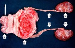 This photograph shows the autopsy specimen from this patient. Included are kidneys (1), ureters (2), bladder (3) (which has been opened), and enlarged prostate (4). Note that the bladder mucosa has multiple trabeculae and the bladder mucosa is hyperemic. Also note that the ureters are dilated.
 -
 This is a close-up of the prostate from this same patient. Note the nodularity of the tissue (1) and the enlargement of the gland. Enlargement of the prostate leads to compression of the urethra as it passes through (2) the gland.
 -
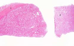 This is a low-power photomicrograph showing hyperplastic prostate on the left (1) and normal prostate on the right (2). At this power, dilated glands are visible in the section of hyperplastic prostate.
 -
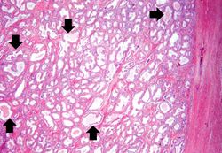 The dilated glands (arrows) make up the major portion of the prostate tissue and there is compression of the stroma.
 -
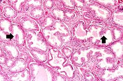 Note these glands, which exhibit hyperplasia of the glandular epithelium. The infolding of the glandular epithelial cells forms papillary projections (arrows) into the lumen of the gland.
 -
 Cystic dilatation of glands is present in this photomicrograph. Notice the accumulation of secretory material inside the glands (arrows) and compression (thinning) of the lining epithelium.
 -
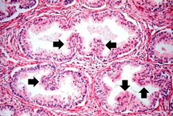 A higher-power view shows the papillary folds (arrows) produced by the hyperplastic epithelium projecting into the lumen of the gland. While these papillary folds project into the lumen of the gland, there is no extension through the glandular basement membrane into the gland's stroma.
 -
 This is a higher-power photomicrograph of papillary folds of hyperplastic epithelium (arrows).
 -
 This kidney was removed from another autopsy patient who had prostatic hyperplasia resulting in marked urinary retention and back-flow of urine from the bladder into the ureters and renal pelvis. The increased pressure inside the renal pelvis resulted in dilation of the renal pelvis (1) and pressure atrophy of the cortex (2). This change in the kidney is called hydronephrosis.


Study Questions
| |||||