IPLab:Lab 12:COPD: Difference between revisions
Jump to navigation
Jump to search
| (One intermediate revision by the same user not shown) | |||
| Line 1: | Line 1: | ||
== Clinical Summary == | == Clinical Summary == | ||
This 64-year-old man was hospitalized because of increasing shortness of breath, cough, increasing sputum production, and fever. The patient had a 75 pack-year history of cigarette smoking. On admission his respiratory rate was 20 breaths per minute and his pulse was 110 bpm. On room air his PaO2 was 46 mm Hg, his PaCO2 was 62 mm Hg, and the pH was 7.26 | This 64-year-old man was hospitalized because of increasing shortness of breath, cough, increasing sputum production, and fever. The patient had a 75 pack-year history of cigarette smoking. On admission his respiratory rate was 20 breaths per minute and his pulse was 110 bpm. On room air his PaO2 was 46 mm Hg, his PaCO2 was 62 mm Hg , and the pH was 7.26. Chest x-ray showed a low, flattened diaphragm, and markedly lucent regions in the upper lung fields suggesting areas of emphysema. Despite appropriate therapy he suffered acute respiratory failure and could not be resuscitated. | ||
Pertinent autopsy findings included emphysema with moderate mucous plugging of bronchi. Right ventricular hypertrophy and dilation were also noted. | |||
Pertinent autopsy findings included emphysema with moderate mucous plugging of bronchi. Right ventricular hypertrophy and dilation were also noted. | |||
== Images == | == Images == | ||
Latest revision as of 21:15, 9 July 2020
Clinical Summary
This 64-year-old man was hospitalized because of increasing shortness of breath, cough, increasing sputum production, and fever. The patient had a 75 pack-year history of cigarette smoking. On admission his respiratory rate was 20 breaths per minute and his pulse was 110 bpm. On room air his PaO2 was 46 mm Hg, his PaCO2 was 62 mm Hg , and the pH was 7.26. Chest x-ray showed a low, flattened diaphragm, and markedly lucent regions in the upper lung fields suggesting areas of emphysema. Despite appropriate therapy he suffered acute respiratory failure and could not be resuscitated.
Pertinent autopsy findings included emphysema with moderate mucous plugging of bronchi. Right ventricular hypertrophy and dilation were also noted.
Images
-
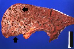 This gross photograph of lung taken at autopsy demonstrates the degree of emphysematous change (arrows). Also note that the rest of the lung is consolidated, indicating a pneumonia.
 -
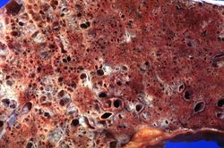 This is a higher-power view of the lung showing the emphysematous change.
 -
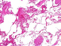 This low-power photomicrograph of the lung demonstrates the enlarged air spaces indicative of emphysematous change.
 -
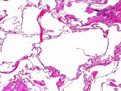 This higher-power photomicrograph of the lung shows more clearly the enlarged air spaces indicative of emphysematous change.
 -
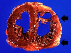 This gross photograph of the heart taken at autopsy demonstrates right ventricular hypertrophy and dilatation (arrows).


Study Questions
- <spoiler text="What is COPD and why do cigarette smokers develop it?">Chronic obstructive pulmonary disease is a group of conditions that lead to dyspnea. COPD entails emphysema, chronic bronchitis, bronchiectasis, and asthma. The conditions can occur to varying degrees and there are often complex combinations and severities of each entity. Cigarette smoke and other air pollutants can irritate the lung and lead to increased leukocytes, increased release of elastases, and increased oxidants which inhibit alpha-1 antitrypsin. These and a variety of other factors lead to destruction of the elastic tissue in the lung and result in abnormal lung function and lung damage.</spoiler>
- <spoiler text="What caused the right ventricular hypertrophy and dilatation? What is this called?">The lung damage resulted in pulmonary hypertension which resulted in right ventricular hypertrophy. Over a period of time the increased workload will cause the right ventricle to fail and dilate. This process of right ventricular hypertrophy due to pulmonary hypertension is called cor pulmonale.</spoiler>
Additional Resources
Reference
- eMedicine Medical Library: Chronic Obstructive Pulmonary Disease and Emphysema in Emergency Medicine
- eMedicine Medical Library: Chronic Obstructive Pulmonary Disease
- eMedicine Medical Library: Nicotine Addiction
- Merck Manual: Chronic Obstructive Pulmonary Disease
Journal Articles
- Franks TJ, Galvin JR. Smoking-Related Interstitial Lung Disease. Arch Pathol Lab Med. 2015;139:974–977.
Images
Related IPLab Cases
| |||||