IPLab:Lab 5:Gaucher Disease: Difference between revisions
Jump to navigation
Jump to search
Seung Park (talk | contribs) No edit summary |
|||
| (6 intermediate revisions by 2 users not shown) | |||
| Line 1: | Line 1: | ||
== Clinical Summary | == Clinical Summary == | ||
This 23-year-old | This 23-year-old female went to her doctor because of chronic fatigue, bone pain, and easy bruising and frequent nose bleeds. Physical exam demonstrated hepatomegaly and splenomegaly and lab results demonstrated anemia and thrombocytopenia. Biopsy and further workup led to a diagnosis of Gaucher disease. | ||
Despite appropriate therapy the anemia and thrombocytopenia persisted as well as the severe splenomegaly. A splenectomy was performed. The surface of the spleen was pale and roughly granular. The cut surface revealed the same pale appearance. | |||
== Images == | == Images == | ||
| Line 16: | Line 15: | ||
File:IPLab5Gaucher8.jpg|This is a higher-power photomicrograph of the spleen from this case. At this higher power individual cells can be better appreciated and the fibrillar nature of the eosinophilic cytoplasmic material can be seen. | File:IPLab5Gaucher8.jpg|This is a higher-power photomicrograph of the spleen from this case. At this higher power individual cells can be better appreciated and the fibrillar nature of the eosinophilic cytoplasmic material can be seen. | ||
</gallery> | </gallery> | ||
== Virtual Microscopy == | |||
<peir-vm>IPLab5Gaucher</peir-vm> | |||
== Study Questions == | == Study Questions == | ||
| Line 30: | Line 32: | ||
* [http://emedicine.medscape.com/article/944157-overview eMedicine Medical Library: Gaucher Disease] | * [http://emedicine.medscape.com/article/944157-overview eMedicine Medical Library: Gaucher Disease] | ||
* [http://www.merckmanuals.com/professional/hematology_and_oncology/spleen_disorders/splenomegaly.html Merck Manual: Splenomegaly] | * [http://www.merckmanuals.com/professional/hematology_and_oncology/spleen_disorders/splenomegaly.html Merck Manual: Splenomegaly] | ||
* [www.merckmanuals.com/professional/pediatrics/inherited_disorders_of_metabolism/lysosomal_storage_disorders.html Merck Manual: Lysosomal Storage Disorders] | * [http://www.merckmanuals.com/professional/pediatrics/inherited_disorders_of_metabolism/lysosomal_storage_disorders.html Merck Manual: Lysosomal Storage Disorders] | ||
=== Journal Articles === | === Journal Articles === | ||
| Line 36: | Line 38: | ||
=== Images === | === Images === | ||
* [ | * [{{SERVER}}/library/index.php?/tags/1670-gaucher_cell PEIR Digital Library: Gaucher Cell Images] | ||
* [http://library.med.utah.edu/WebPath/HEMEHTML/HEMEIDX.html Webpath: Hematopathology] | * [http://library.med.utah.edu/WebPath/HEMEHTML/HEMEIDX.html Webpath: Hematopathology] | ||
Latest revision as of 14:52, 8 July 2020
Clinical Summary
This 23-year-old female went to her doctor because of chronic fatigue, bone pain, and easy bruising and frequent nose bleeds. Physical exam demonstrated hepatomegaly and splenomegaly and lab results demonstrated anemia and thrombocytopenia. Biopsy and further workup led to a diagnosis of Gaucher disease.
Despite appropriate therapy the anemia and thrombocytopenia persisted as well as the severe splenomegaly. A splenectomy was performed. The surface of the spleen was pale and roughly granular. The cut surface revealed the same pale appearance.
Images
-
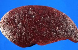 This is a gross photograph of spleen from this case. The spleen is enlarged and the surface is finely granular.
 -
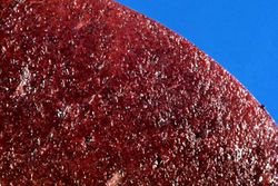 This is a cut section of spleen from this case. Again note the fine granular appearance to the tissue.
 -
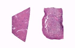 This is a low-power photomicrograph of normal spleen (left) and the spleen from this case (right). The loose appearance of the tissue in the Gaucher spleen is due to artifactual loss of tissue during histologic processing.
 -
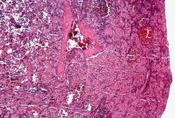 This is a photomicrograph of the spleen from this case. There is very little if any white pulp evident in this section.
 -
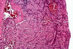 This is a higher-power photomicrograph of the spleen from this case. Again there is no white pulp and the red pulp is filled with large eosinophilic cells.
 -
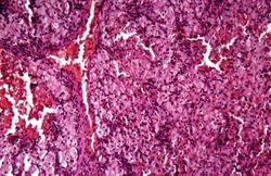 This is another high-power photomicrograph of the spleen from this case. At this power it is easier to see the large eosinophilic cells.
 -
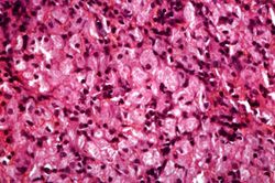 This is another high-power photomicrograph of the spleen from this case. At this high power individual cells can be better appreciated.
 -
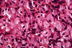 This is a higher-power photomicrograph of the spleen from this case. At this higher power individual cells can be better appreciated and the fibrillar nature of the eosinophilic cytoplasmic material can be seen.


Virtual Microscopy
Study Questions
Additional Resources
Reference
- eMedicine Medical Library: Gaucher Disease
- Merck Manual: Splenomegaly
- Merck Manual: Lysosomal Storage Disorders
Journal Articles
- Barone R, Pavone V, Nigro F, Chabàs A, Fiumara A. Extraordinary bone involvement in a gaucher disease type I patient. Br J Haematol 2000 Mar;108(4):838-41.
Images
| |||||