Difference between revisions of "IPLab:Lab 12:Radiation Changes"
Seung Park (talk | contribs) |
Seung Park (talk | contribs) |
||
| Line 16: | Line 16: | ||
File:IPLab12RadiationChanges8.jpg|This high-power photomicrograph of the wall of the ileum shows more examples of pleomorphic cells caused by radiation injury (arrows). | File:IPLab12RadiationChanges8.jpg|This high-power photomicrograph of the wall of the ileum shows more examples of pleomorphic cells caused by radiation injury (arrows). | ||
</gallery> | </gallery> | ||
| + | |||
| + | == Study Questions == | ||
| + | * <spoiler text="Why was there an obstruction at this site?">Radiation damage caused fibrosis of the bowel wall and atrophy of the mucosa. This probably led to an area of poor peristalsis and a physiologic obstruction. The fibrosis in the wall can also lead to a physical obstruction (stricture). | ||
| + | |||
| + | Due to the atrophy of the mucosa, the fibrosis, and the poor blood flow to this area (chronic vascular occlusive lesions), this area of bowel would be more susceptible to rupture, as happened in this case.</spoiler> | ||
| + | * <spoiler text="Why are the small intestine and colon so susceptible to radiation injury?">The gastrointestinal tract is very radiosensitive and is frequently affected in all forms of deep radiation. The intestinal epithelium (crypt cells) are particularly radiosensitive due to their high turnover rate. These cell often show nuclear and cellular pleomorphism, mitotic abnormalities, and cell necrosis. Later, ulcerations may appear, along with vascular and connective tissue changes. The late effects of intestinal injury comprise mucosal and submucosal atrophy and fibrosis, occasionally producing intestinal strictures.</spoiler> | ||
| + | * <spoiler text="Are patients who have been treated with radiation more susceptible to formation of new cancers?">Yes, due to the above-mentioned alterations in nuclear and mitotic activity.</spoiler> | ||
{{IPLab 12}} | {{IPLab 12}} | ||
[[Category: IPLab:Lab 12]] | [[Category: IPLab:Lab 12]] | ||
Revision as of 16:27, 21 August 2013
Clinical Summary[edit]
This 46-year-old white female was found to have prolapse of the uterus two years earlier for which a vaginal hysterectomy had been performed. Study of the specimen demonstrated invasive squamous cell carcinoma of the cervix. Subsequently, she underwent a radical parametrectomy, removal of both tubes and ovaries, and partial resection of the bladder. Six months later, a recurrence of the tumor was treated by 6000 rads to the whole pelvis. Subsequently, the patient developed intermittent small bowel obstruction which resulted in the resection of a segment of ileum. She did well following surgery.
Autopsy Findings[edit]
The surgical specimen consisted of a 13-cm segment of ileum having a luminal circumference of 5 cm. There was a full-thickness tear measuring 2.5 cm in the center of the specimen. On opening the bowel, the mucosa was ulcerated and showed a perforation.
Images[edit]
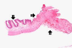
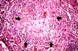
This is a low-power photomicrograph of the surgical specimen of the ileum. The normal ileum is to the left (1). The area of stricture consists of dense fibrous connective tissue (2) and there is loss or marked atrophy of the epithelium (3).
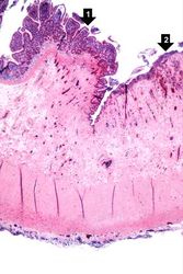
This is a higher-power photomicrograph of the surgical specimen of the ileum showing the transition from the normal epithelium (1) to the atrophied epithelium (2) in the area of radiation injury.
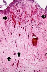
This is a higher-power photomicrograph showing the atrophied epithelium in the area of radiation injury. There are some epithelial cells deep within the mucosa (1). Note the dense fibrous connective tissue (2) within the wall of the ileum.
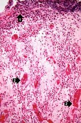
This is a high-power photomicrograph showing the atrophied epithelium in the area of radiation injury (1). Note the dense fibrous connective tissue within the wall of the ileum and the congested blood vessels (2).
This is a high-power photomicrograph of the wall of the ileum showing a blood vessel that has suffered radiation-induced damage and is completely occluded (arrows).
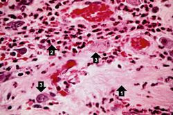
This high-power photomicrograph of the wall of the ileum shows areas of fibrosis (1), inflammatory cells (2), and abnormal pleomorphic cells (3) in the area of radiation injury. The abnormal morphology of these cells is radiation-induced. These cells are often difficult to distinguish from recurrent tumor cells.
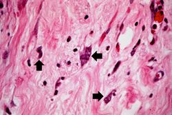
This high-power photomicrograph of the wall of the ileum shows more examples of pleomorphic cells (arrows) due to radiation injury.
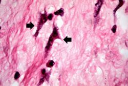
This high-power photomicrograph of the wall of the ileum shows more examples of pleomorphic cells caused by radiation injury (arrows).


Study Questions[edit]
| |||||
Parametrectomy is the surgical removal of supporting tissues that surround the uterus.