IPLab:Lab 9:Actinomycosis
Jump to navigation
Jump to search
Clinical Summary
This 18-year-old black female felt well until one year before death, when she developed a persistent, progressive skin rash and weight loss. One month before death, draining abscesses appeared in the perirectal region. Biopsy showed actinomycosis. Despite treatment, the patient died.
Autopsy revealed a large abscess around the cecum which had ruptured. The perirectal abscesses had originated from extensions of this pericecal abscess.
Images
-
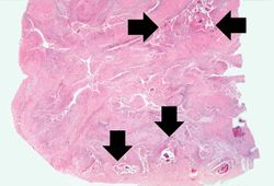 This is a low-power photomicrograph of the retroperitoneal abscess. At this magnification, multiple dark-staining foci can be appreciated. These foci are Actinomyces colonies (arrows). These colonies are known as "sulfur granules" because in gross specimens they are visible to the naked eye as yellow grains, thus resembling grains of sulfur.
 -
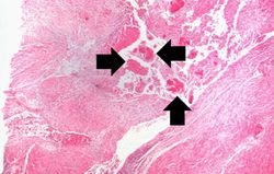 This is a higher-power photomicrograph of an abscess demonstrating a pocket of purulent exudate that contains numerous actinomycotic colonies (arrows).
 -
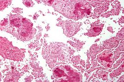 This is a higher-power photomicrograph of actinomycotic colonies in the abscess.
 -
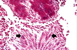 This is an even higher-power photomicrograph of actinomycotic colonies in the abscess. The filamentous nature (arrows) of the actinomyces organisms in these colonies can be appreciated.
 -
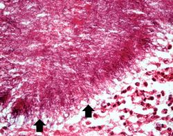 This is a high-power photomicrograph of an actinomycotic colony. The filamentous nature (arrows) of the actinomyces organisms is more easily appreciated at this power.


Virtual Microscopy
Study Questions
Additional Resources
Reference
- eMedicine Medical Library: Actinomycosis
- eMedicine Medical Library: Pediatric Actinomycosis
- Merck Manual: Actinomycosis
Journal Articles
- Yildiz O, Doganay M. Actinomycoses and Nocardia pulmonary infections. Curr Opin Pulm Med 2006 May;12(3):228-34.
Images
| |||||