Difference between revisions of "IPLab:Lab 9:Diphtheria"
Seung Park (talk | contribs) |
Seung Park (talk | contribs) |
||
| Line 12: | Line 12: | ||
File:IPLab9Diphtheria4.jpg|In this higher-power photomicrograph of the tissue from the previous image, the ulcerated tracheal mucosa and the diphtheritic membrane are more clearly seen. Although difficult to make out at this magnification, most of the cells in this inflammatory exudate are neutrophils. | File:IPLab9Diphtheria4.jpg|In this higher-power photomicrograph of the tissue from the previous image, the ulcerated tracheal mucosa and the diphtheritic membrane are more clearly seen. Although difficult to make out at this magnification, most of the cells in this inflammatory exudate are neutrophils. | ||
</gallery> | </gallery> | ||
| + | |||
| + | == Study Questions == | ||
| + | * <spoiler text="What organism causes diphtheria?">Diphtheria is caused by Corynebacterium diphtheriae. This organism is passed from person to person by aerosols or by skin shedding.</spoiler> | ||
| + | * <spoiler text="What toxins are involved in the pathogenesis of diphtheria?">C. diphtheriae has only one toxin, which is encoded by a lysogenic phage. Thus, C. diphtheriae cannot cause diphtheria unless the phage first infects the bacteria and provides it with the DNA necessary for production of the toxin.</spoiler> | ||
| + | * <spoiler text="How does diphtheria toxin cause cell killing?">The toxin is composed of fragment B that attaches to host cells and fragment A which is linked to fragment B by a disulfide bridge. Bound diphtheria toxin enters the acidic endosome of cells, fuses with the endosomal membrane, and then enters the cell cytoplasm. There the disulfide bond of the toxin is broken, releasing the enzymatically active fragment A which then catalyzes the covalent transfer of adenosine diphosphate ribose (ADPR) from nicotinamide-adenine dinucleotide (NAD) to EF-2. EF-2, a ribosomal elongation factor necessary for protein synthesis, is thus inactivated. One molecule of diphtheria toxin can kill a cell by ADP-ribosylating more than a million EF-2 molecules.</spoiler> | ||
{{IPLab 9}} | {{IPLab 9}} | ||
[[Category: IPLab:Lab 9]] | [[Category: IPLab:Lab 9]] | ||
Revision as of 15:56, 21 August 2013
Clinical Summary[edit]
This 4-year-old black female had an upper respiratory infection and a sore throat with increasing difficulty in breathing. Membranous exudate over one tonsil led to a working diagnosis of diphtheria, and the child was admitted. On the day of her admission, the child developed signs of respiratory tract obstruction and a tracheotomy was performed. However, the procedure was unable to establish a patent airway and the child died.
Autopsy Findings[edit]
At autopsy, a dense grayish pink membrane extended from both tonsils to the mid-trachea. The lungs were edematous and showed signs of pneumonia.
Images[edit]
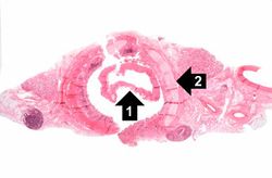
This is a low-power photomicrograph of the trachea with the diphtheritic membrane (1), which has pulled away from the tracheal lining during histological processing. Note the tracheal cartilage (2) present in this section.
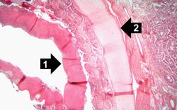
This is a higher-power photomicrograph of trachea with the diphtheritic membrane (1). Even though, the main part of the membrane has pulled away from the tracheal lining during histological processing, in this section part of the membrane is still loosely attached. Once again, note the tracheal cartilage (2).
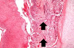
This is an even higher-power photomicrograph of the tracheal mucosa and the diphtheritic membrane. The mucosal surface of the trachea is ulcerated (total loss of epithelial cells) and the only remaining epithelial cells are found in the glands (arrows). The diphtheritic membrane consists of fibrin and inflammatory cells, most of which are dead.
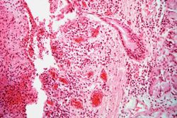
In this higher-power photomicrograph of the tissue from the previous image, the ulcerated tracheal mucosa and the diphtheritic membrane are more clearly seen. Although difficult to make out at this magnification, most of the cells in this inflammatory exudate are neutrophils.


Study Questions[edit]
| |||||
In alcoholics, aspiration pneumonia is common--bacteria enter the lung via aspiration of gastric contents.