Difference between revisions of "IPLab:Lab 9:Diphtheria"
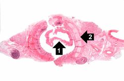
Seung Park (talk | contribs) (Created page with "== Images == <gallery heights="250px" widths="250px"> File:IPLab9Diphtheria1.jpg|This is a low-power photomicrograph of the trachea with the diphtheritic membrane (1), which h...") |
Seung Park (talk | contribs) |
||
| Line 1: | Line 1: | ||
| + | == Clinical Summary == | ||
| + | This 4-year-old black female had an upper respiratory infection and a sore throat with increasing difficulty in breathing. Membranous exudate over one tonsil led to a working diagnosis of diphtheria, and the child was admitted. On the day of her admission, the child developed signs of respiratory tract obstruction and a tracheotomy was performed. However, the procedure was unable to establish a patent airway and the child died. | ||
| + | |||
| + | == Autopsy Findings == | ||
| + | At autopsy, a dense grayish pink membrane extended from both tonsils to the mid-trachea. The lungs were edematous and showed signs of pneumonia. | ||
| + | |||
== Images == | == Images == | ||
<gallery heights="250px" widths="250px"> | <gallery heights="250px" widths="250px"> | ||
Revision as of 14:37, 21 August 2013
Clinical Summary[edit]
This 4-year-old black female had an upper respiratory infection and a sore throat with increasing difficulty in breathing. Membranous exudate over one tonsil led to a working diagnosis of diphtheria, and the child was admitted. On the day of her admission, the child developed signs of respiratory tract obstruction and a tracheotomy was performed. However, the procedure was unable to establish a patent airway and the child died.
Autopsy Findings[edit]
At autopsy, a dense grayish pink membrane extended from both tonsils to the mid-trachea. The lungs were edematous and showed signs of pneumonia.
Images[edit]
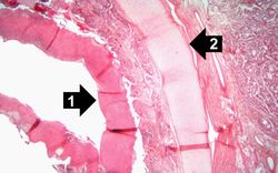
This is a higher-power photomicrograph of trachea with the diphtheritic membrane (1). Even though, the main part of the membrane has pulled away from the tracheal lining during histological processing, in this section part of the membrane is still loosely attached. Once again, note the tracheal cartilage (2).
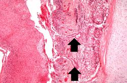
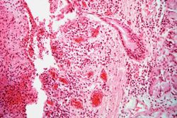
This is an even higher-power photomicrograph of the tracheal mucosa and the diphtheritic membrane. The mucosal surface of the trachea is ulcerated (total loss of epithelial cells) and the only remaining epithelial cells are found in the glands (arrows). The diphtheritic membrane consists of fibrin and inflammatory cells, most of which are dead.
| |||||
In alcoholics, aspiration pneumonia is common--bacteria enter the lung via aspiration of gastric contents.


