Difference between revisions of "IPLab:Lab 9:Clostridial Myonecrosis"
Seung Park (talk | contribs) |
Seung Park (talk | contribs) |
||
| Line 12: | Line 12: | ||
File:IPLab9Clostridium7.jpg|This is a high-power photomicrograph of a tissue section stained with a tissue Gram's stain (Brown & Brenn). The Gram-positive bacilli can be seen throughout this tissue section. | File:IPLab9Clostridium7.jpg|This is a high-power photomicrograph of a tissue section stained with a tissue Gram's stain (Brown & Brenn). The Gram-positive bacilli can be seen throughout this tissue section. | ||
</gallery> | </gallery> | ||
| + | |||
| + | == Study Questions == | ||
| + | * <spoiler text="What organism causes myonecrosis (gas gangrene)?">Clostridium perfringens invades traumatic or surgical wounds and causes an anaerobic cellulitis or myonecrosis (gas gangrene). Clostridium species are gram-positive bacilli that grow under anaerobic conditions and produce spores which are frequently present in the soil.</spoiler> | ||
| + | * <spoiler text="What are some of the factors that help Clostridium perfringens produce such a fulminant anaerobic cellulitis?">Clostridia release collagenase and hyaluronidase that degrade the extracellular matrix proteins (collagen and elastin) which make up the connective tissue between muscle bundles. This allows the bacteria to migrate and invade along fascial planes. | ||
| + | |||
| + | C. perfringens secrete 12 toxins, the most important of which is alpha toxin. Alpha toxin is a phospholipase C that degrades lecithin, a major component of cell membranes, and thus destroys red blood cells, platelets, and muscle cells, causing myonecrosis. | ||
| + | |||
| + | Alpha toxin also has a sphingomyelinase activity that contributes to nerve-sheath damage. | ||
| + | |||
| + | Theta toxin binds cholesterol and forms a membrane-destabilizing pore that causes leukocyte lysis, explaining the paucity of polymorphonuclear leukocytes in the lesions of gas gangrene.</spoiler> | ||
{{IPLab 9}} | {{IPLab 9}} | ||
[[Category: IPLab:Lab 9]] | [[Category: IPLab:Lab 9]] | ||
Revision as of 15:59, 21 August 2013
Clinical Summary[edit]
This 68-year-old white male with insulin-dependent diabetes was admitted one day before his death. The chief complaints were the occurrence of chills and fever since passing a kidney stone two days earlier. In the last day, the right leg had become swollen. The most striking physical findings were redness of the right posterior calf and crepitance in both legs. The patient's white blood cell count was found to be 34,000 cells/cmm and the packed red blood cell volume (PCV) was 18%. Within hours, the right calf became tense and the crepitance spread up to the nipple line. The patient vomited, aspirated the vomitus, and died 10 hours after admission.
Images[edit]
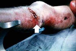
This gross photograph of the lower extremity was taken at autopsy. Notice the swelling and the area of the primary infection (arrow).
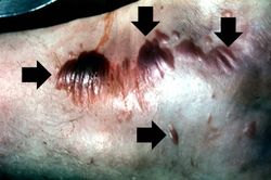
This gross photograph shows a close-up view of hemorrhagic blebs (arrows) on the skin. The blebs on the skin are accumulations of gas being discharged into the tissues from the Clostridium perfringens. This gas produces crepitance.
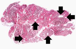
This is a low-power photomicrograph of muscle fascicles containing large gas bubbles (arrows). Note that there is no inflammatory reaction in this section.
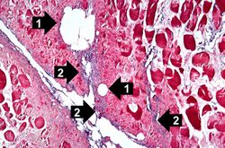
This is a high-power photomicrograph of skeletal muscle. The muscle cells are hypereosinophilic and most do not contain nuclei, indicating that these cells are dead or dying. The round clear spaces (1) in this tissue correspond to gas accumulations prior to death. In between the bundles of muscle cells, accumulations of small dark blue-staining bacterial organisms can be seen (2). Also note that there is no inflammatory response in this tissue.
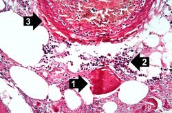
This high-power photomicrograph shows the gas accumulation present in the tissue, a necrotic muscle cell (1), and a mild inflammatory response (2). There is also a thrombosed blood vessel (3). The blue-staining rods (bacterial organisms) can barely be appreciated at this magnification.
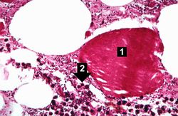
This higher-power photomicrograph of the previous image provides a clearer view of gas bubbles in the tissue, the necrotic hypereosinophilic muscle cell (1), and the mild inflammatory reaction (2). At this magnification, the bacteria located throughout this section can be better appreciated.
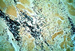
This is a high-power photomicrograph of a tissue section stained with a tissue Gram's stain (Brown & Brenn). The Gram-positive bacilli can be seen throughout this tissue section.


Study Questions[edit]
| |||||
A normal hematocrit for a male is 39 to 49%.