Difference between revisions of "IPLab:Lab 3:Chronic Peptic Ulcer"
(→Journal Articles) |
(→Images) |
||
| (2 intermediate revisions by the same user not shown) | |||
| Line 1: | Line 1: | ||
== Clinical Summary == | == Clinical Summary == | ||
| − | + | A 56-year-old white female was admitted to the hospital with severe epigastric pain and burning. This pain, which has occurred intermittently for the Last 4 years was said to be worse at night and on an empty stomach. She had also experienced several episodes of hematemesis and melena. An upper endoscopy was performed which demonstrated an ulcer in the gastric mucosa. A breath test for Helicobacter pylori was positive so the patient was started on antibiotics and proton pump inhibitors (PPI). | |
| + | |||
| + | Autopsy pictures and histology sections from a different patient with peptic ulcer disease are presented. | ||
== Images == | == Images == | ||
<gallery heights="250px" widths="250px"> | <gallery heights="250px" widths="250px"> | ||
| − | File: | + | File:IPLab3ChronicPepticUlcer1b.jpg|This is a gross photograph of a stomach containing two ulcers. Note the gastric mucosa that extends up to the edge of the ulcer (arrows). |
File:IPLab3ChronicPepticUlcer2.jpg|This is a gross photograph of the ulcer after it has been transected. The edge of the mucosa (1) is better appreciated in this image. Note the thick, fatty tissue (2) which makes up the base of this ulcer (3). | File:IPLab3ChronicPepticUlcer2.jpg|This is a gross photograph of the ulcer after it has been transected. The edge of the mucosa (1) is better appreciated in this image. Note the thick, fatty tissue (2) which makes up the base of this ulcer (3). | ||
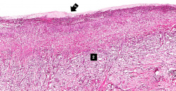
| − | File: | + | File:IPLab3ChronicPepticUlcer3b.JPG|This is a low-power photomicrograph of a section through the transected ulcer. The blue cells on the right hand side of this section are the normal gastric epithelial cells of the mucosa (1). Note the absence of any epithelial cells within the crater of the ulcer (2). |
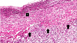
| − | File: | + | File:IPLab3ChronicPepticUlcer4b.JPG|This is a photomicrograph of the margin of the ulcer. Note the intact epithelium on the right side of the section (1) and the ulcerated region without epithelium on the left (2). There are numerous inflammatory cells within this tissue. |
| − | File: | + | File:IPLab3ChronicPepticUlcer5b.JPG|This is a medium-power photomicrograph of the base of the ulcer with the fibrinopurulent membrane (1) overlying the ulcerated surface. The ulcerated surface contains granulation tissue and inflammatory cells (2). |
| − | File: | + | File:IPLab3ChronicPepticUlcer6b.JPG|This high-power photomicrograph of the ulcer base (arrows) demonstrates the lack of epithelium and the exuberant inflammatory response (1) consisting of primarily of fibrin and neutrophils (PMNs). The surface of the ulcer bed is covered with this fibrinopurulent exudate. |
| − | File: | + | File:IPLab3ChronicPepticUlcer7b.JPG|This high-power photomicrograph of the ulcer base demonstrates plump, activated fibroblasts and endothelial cells (arrows) within the granulation tissue that makes up the base of the ulcer. There are inflammatory cells (primarily lymphocytes) within this region as well. |
| − | File: | + | File:IPLab3ChronicPepticUlcer8b.JPG|This low-power photomicrograph demonstrates the healing reaction in the base of this ulcer. The base of the ulcer is at the top of the image and the serosal surface is at the bottom. Note the granulation tissue and fibrous connective tissue within the wall of the stomach (1) and the layer of inflammatory exudate on the surface of the ulcer (arrow). |
| − | |||
| − | |||
| − | |||
</gallery> | </gallery> | ||
Latest revision as of 00:09, 20 June 2020
Contents
Clinical Summary[edit]
A 56-year-old white female was admitted to the hospital with severe epigastric pain and burning. This pain, which has occurred intermittently for the Last 4 years was said to be worse at night and on an empty stomach. She had also experienced several episodes of hematemesis and melena. An upper endoscopy was performed which demonstrated an ulcer in the gastric mucosa. A breath test for Helicobacter pylori was positive so the patient was started on antibiotics and proton pump inhibitors (PPI).
Autopsy pictures and histology sections from a different patient with peptic ulcer disease are presented.
Images[edit]
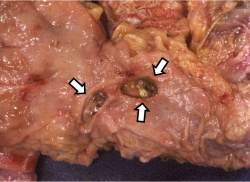
This is a gross photograph of a stomach containing two ulcers. Note the gastric mucosa that extends up to the edge of the ulcer (arrows).
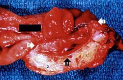
This is a gross photograph of the ulcer after it has been transected. The edge of the mucosa (1) is better appreciated in this image. Note the thick, fatty tissue (2) which makes up the base of this ulcer (3).

This is a low-power photomicrograph of a section through the transected ulcer. The blue cells on the right hand side of this section are the normal gastric epithelial cells of the mucosa (1). Note the absence of any epithelial cells within the crater of the ulcer (2).

This is a photomicrograph of the margin of the ulcer. Note the intact epithelium on the right side of the section (1) and the ulcerated region without epithelium on the left (2). There are numerous inflammatory cells within this tissue.
This is a medium-power photomicrograph of the base of the ulcer with the fibrinopurulent membrane (1) overlying the ulcerated surface. The ulcerated surface contains granulation tissue and inflammatory cells (2).
This high-power photomicrograph of the ulcer base (arrows) demonstrates the lack of epithelium and the exuberant inflammatory response (1) consisting of primarily of fibrin and neutrophils (PMNs). The surface of the ulcer bed is covered with this fibrinopurulent exudate.
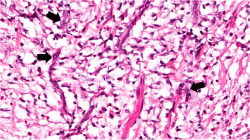
This high-power photomicrograph of the ulcer base demonstrates plump, activated fibroblasts and endothelial cells (arrows) within the granulation tissue that makes up the base of the ulcer. There are inflammatory cells (primarily lymphocytes) within this region as well.

This low-power photomicrograph demonstrates the healing reaction in the base of this ulcer. The base of the ulcer is at the top of the image and the serosal surface is at the bottom. Note the granulation tissue and fibrous connective tissue within the wall of the stomach (1) and the layer of inflammatory exudate on the surface of the ulcer (arrow).


Virtual Microscopy[edit]
Study Questions[edit]
Additional Resources[edit]
Reference[edit]
Journal Articles[edit]
- Laine L. Upper Gastrointestinal Bleeding Due to a Peptic Ulcer. NEJM 2016 June 22; 374: 2367-2376.
Images[edit]
Hematemesis is the vomiting of blood.
Melena is the passage of digested blood in the feces.
Cirrhosis is a liver disease characterized by necrosis, fibrosis, loss of normal liver architecture, and hyperplastic nodules.
Hypercalcemia is the state of having increased levels of calcium in the blood.
The normal fibrinogen level is 184 to 412 mg/dL.