IPLab:Lab 9:Bacterial Meningitis
Clinical Summary
This 45-year-old white female with a history of psychiatric illness sustained self-inflicted third degree burns over 49% of her body surface. After initially doing well under treatment, she developed severe respiratory distress and became comatose. Antemortem blood cultures were positive for Pseudomonas aeruginosa.
Images
-
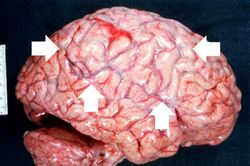 This gross photograph of the autopsy specimen from this case demonstrates the purulent exudate (arrows) in the leptomeninges.
 -
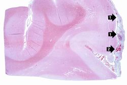 This is a low-power photomicrograph of brain section. Note the exudate (1) in the meninges and congestion of the vessels (2) in the leptomeninges.
 -
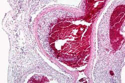 This is a higher-power view of a congested blood vessel. Inflammatory exudate is present within the vessel and throughout the leptomeninges.
 -
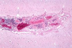 This higher-power photomicrograph of a sulcus shows the congested vessels and the inflammatory exudate in the leptomeninges.
 -
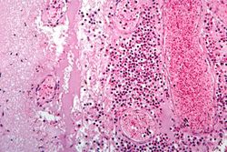 This is a higher-power photomicrograph of inflammatory exudate in a sulcus. The majority of cells in this exudate are neutrophils. There is also abundant fibrin (arrows) and red blood cells are present in the congested vessels.
 -
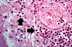 This is a high-power photomicrograph of a blood vessel from the previous image. The vessel is surrounded by neutrophils (arrows).
 -
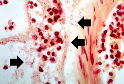 This is a high-power photomicrograph of exudate from the leptomeninges which has been Gram-stained. Note the Gram-negative bacteria (arrows) throughout this section.
 -
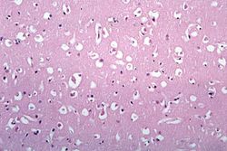 This photomicrograph of brain tissue demonstrates diffuse edema.


Virtual Microscopy
Study Questions
Additional Resources
Reference
- eMedicine Medical Library: Pediatric Bacterial Meningitis
- eMedicine Medical Library: Meningitis
- eMedicine Medical Library: Thermal Burns
- eMedicine Medical Library: Pseudomonas aeruginosa Infections
- Merck Manual: Acute Bacterial Meningitis
- Merck Manual: Burns
Journal Articles
- Grände PO, Myhre EB, Nordström CH, Schliamser S. Treatment of intracranial hypertension and aspects on lumbar dural puncture in severe bacterial meningitis. Acta Anaesthesiol Scand 2002 Mar;46(3):264-70.
Images
| |||||